. 2014 Feb 27;9(2):e89983.
doi: 10.1371/journal.pone.0089983. eCollection 2014.

Deciphering the stromal and hematopoietic cell network of the adventitia from non-aneurysmal and aneurysmal human aorta

Affiliations

Deciphering the stromal and hematopoietic cell network of the adventitia from non-aneurysmal and aneurysmal human aorta

Charles-Antoine Dutertre et al. PLoS One. .

Abstract

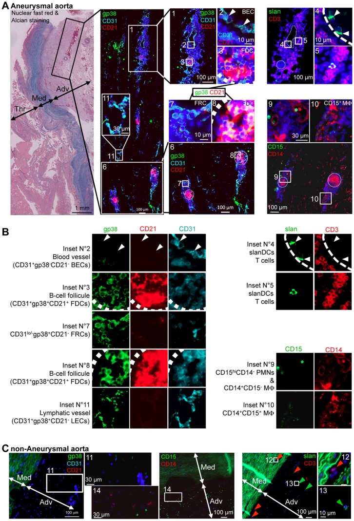
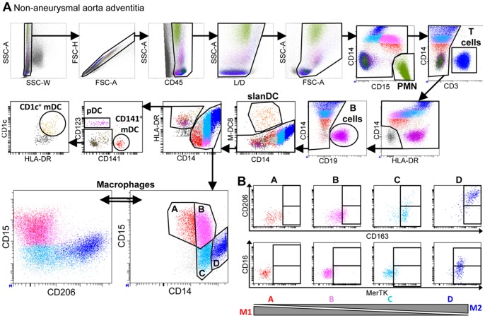
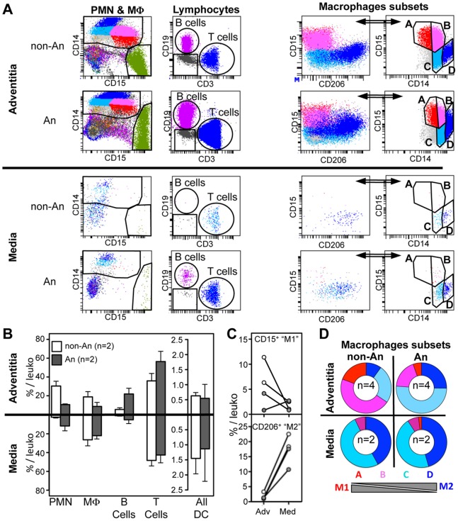
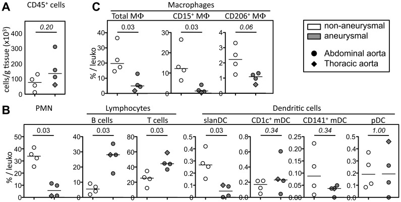
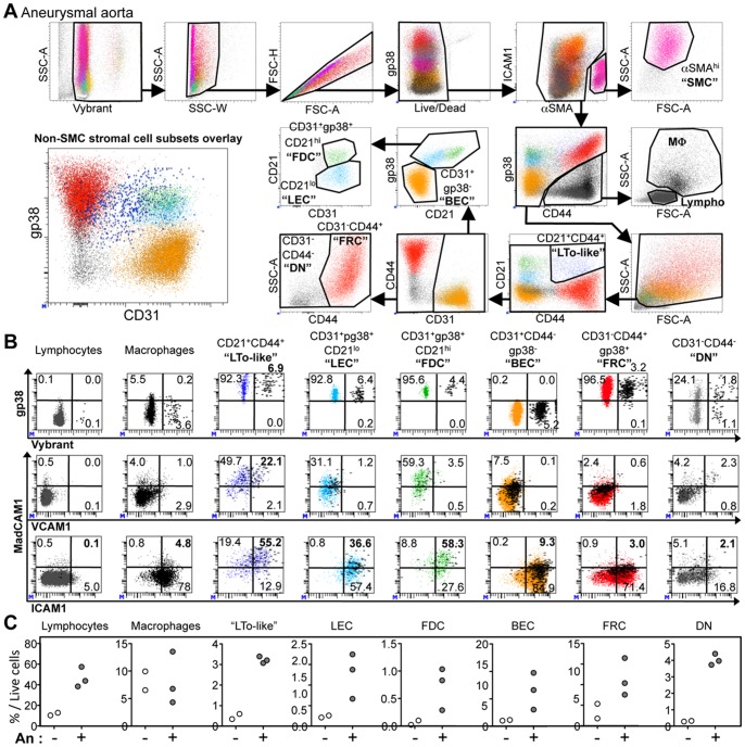
Aneurysm is associated to a complex remodeling of arteries that affects all their layers. Although events taking place in the intima and the media have received a particular attention, molecular and cellular events taking place in the adventitia have started to be deciphered only recently. In this study, we have precisely described the composition and distribution of stromal and hematopoietic cells in human arterial adventitia, both at steady state and in the setting of aortic aneurysm. Using polychromatic immunofluorescent and flow cytometry analyses, we observed that unlike the medial layer (which comprises mostly macrophages and T cells among leukocytes), the adventitia comprises a much greater variety of leukocytes. We observed an altered balance in macrophages subsets in favor of M2-like macrophages, an increased proliferation of macrophages, a greater number of all stromal cells in aneurysmal aortas. We also confirmed that in this pathological setting, adventitia comprised blood vessels and arterial tertiary lymphoid organs (ATLOs), which contained also M-DC8(+) dendritic cells (slanDCs) that could participate in the induction of T-cell responses. Finally, we showed that lymphatic vessels can be detected in aneurysmal adventitia, the functionality of which will have to be evaluated in future studies. All together, these observations provide an integrative outlook of the stromal and hematopoietic cell network of the human adventitia both at steady state and in the context of aneurysm.


Conflict of interest statement

Competing Interests: The authors have declared that no competing interests exist.

Figures

Figure 1
Figure 1. Blood and lymphatic vessels are found in the adventitia of aneurysmal aortas, which also comprises lymphoid structures composed of stromal cells and leukocytes.
Reresentative immunohistofluorescence analyses of paraffin-embedded (A) aneurysmal or (B) non-aneurysmal aortas. (A) The far left panels shows a micrograph of a nuclear fast red and Alcian blue-stained section. The left immunofluorescent cross-section micrograph corresponds to gp38 (green), CD31 (cyan) and CD21 (red) staining to study stromal cell subsets localization, and corresponds to the region defined by a black rectangle in the Nuclear fast red & Alcian micrograph. Magnifications of regions defined by white rectangles are shown as insets and in the two middle panels, numbered white boxes indicate the position of the magnified insets, and cell subsets defined in insets are sometimes specified. In each middle panel inset, two out of the three staining (CD31 and CD21 in the upper inset, or gp38 and CD21 for the other insets) are shown. Sequential sections following the one stained for gp38/CD31/CD21 were stained for slan (M-DC8, green) and CD3 (red) to detect slanDC and CD3+ T cells (upper right panels), or for CD15 (green) and CD14 (red) to detect macrophages (CD14+CD15+/−) and polymorphonuclear cells (CD15+CD14) (lower right panels). Numbered white boxes indicate the position of the magnified insets. (B) Single channels for insets defined in (A) are show. The cell types and/or structure defined are written on the left of each inset. (C) Sequential sections of a paraffin embedded non-aneurysmal aorta were stained for gp38 (green), CD31 (cyan) and CD21 (red) (left panels), for slan (M-DC8, green) and CD3 (red) (middle panels), or for CD15 (green) and CD14 (red) (right panels). Numbered white boxes indicate the position of the magnified insets. Thr, thrombus; Med, media; Adv, adventitia.
Figure 2
Figure 2. Analysis of leukocyte subsets in the adventitia from a non-aneurysmal abdominal aorta by polychromatic flow cytometry.
(A) The gating strategy to analyze CD45+ leukocytes from a non-aneurysmal abdominal aorta by 15-color flow cytometry is shown. This strategy allows the simultaneous definition of CD14CD15hi polymorphonuclear cells (PMN), CD3+ T cells, CD19+ B cells, CD14+ macrophages, and dendritic cell DC) subsets comprising CD14loM-DC8+ slanDC, and among linHLA-DR+ events, CD123+ plasmacytoid DC (pDC), and two subsets of myeloid DC (mDC), CD141+ mDC and CD1c+ mDC. The lower right dot plot shows four macrophage subsets defined by their relative expression of CD14 and CD15, and the lower left panel shows CD206 and CD15 expression by these macrophage subsets. (B) Flow cytometric dot plots of CD163 and CD206 (upper dot plots) and MerTK and CD16 (lower dot plots) expression by the four macrophage subsets defined in (A).
Figure 3
Figure 3. Comparison of leukocyte subsets in the adventitia and the media from non-aneurysmal and aneurysmal abdominal aortas.
(A) Flow cytometric dot plots defining polymorphonuclear cells (PMN) and macrophages (MΦ, far left dot plots), CD3+ T cells and CD19+ B cells (middle left dot plots), and macrophages subsets (right dot plots) in the adventitia (upper panels) and the media (lower panels) from a non-aneurysmal (non-An) and an aneurysmal (An) abdominal aorta. (B) On the left, graphical representation of the percentages among total CD45+ leukocytes of PMN, macrophages, B cells, T cells and all DC in the adventitia (n = 2, upper histograms), and the media (n = 2, lower histograms) from non-An (white histograms) and An (grey histograms) aortas. (C) Comparison between the adventitia (Adv) and the media (Med) for the percentages among total CD45+ leukocytes of CD15+ and CD206+ macrophage subsets in non-An (white filled circles) and An (grey filled circles) aortas (lines associates paired results obtained in the adventitia and the media from the same patient). (D) Ring graphical representation of the proportion of the macrophage subsets defined in Figure 2A among total macrophages in the adventitia (n = 4, top panels) and the media (n = 2, bottom panels).
Figure 4
Figure 4. Proportion of leukocyte subsets in the adventitia of non-aneurysmal and aneurysmal aortas.
(A) Graphical representation of the density of CD45+ leukocytes (cells/g tissue), and of (B,C) the percentage among CD45+ leukocytes of (B) PMN, lymphocyte, DC, and (C) macrophages and their subsets in the adventitia from non-aneurysmal (white filled symbols, n = 4) and aneurysmal (grey filled symbols, n = 4) aortas. Circles correspond to abdominal aortas, and the diamond to a thoracic aorta. p values were calculated with the Mann-Whitney nonparametric test.
Figure 5
Figure 5. Analysis of stromal cell subsets in the adventitia form non-aneurysmal and aneurysmal aortas.
(A) The gating strategy to analyze all nucleated cells (Vybrant+) and particularly stromal cell subsets from an aneurysmal artery by 10-color flow cytometry is shown. This strategy allows the simultaneous definition of residual aSMAhi smooth muscle cells (SMC), CD44+gp38lo/− leukocytes [macrophages (MΦ) and lymphocytes (Lympho) defined on the forward (FSC-A) and side (SSC-A) scatter], among non-leukocyte cells, CD21+CD44+ “LTo-like” cells, CD31+CD44pg38 blood endothelial cells (BEC), CD31+gp38+CD21lo lymphatic endothelial cells (LEC), CD31+gp38+CD21hi follicular dendritic cells (FDC), CD31CD44+ fibroblastic reticular cells (FRC), and CD31CD44 double negative cells (DN). The lower left dot plot (CD31 vs pg38) shows the overlay of all stromal cell subsets excluding SMC. (B) Flow cytometric dot plots showing the expression of Vybrant (Vybranthi cells being undergoing division) and gp38 (upper panels), VCAM1 and MadCAM1 (middle panels) or ICAM1 and MadCAM1 (lower panels) for cell subsets defined in (A). The percentage of cells in each quadrant is indicated. Vybrant-positive events are highlighted and displayed as thick black dots in the middle and lower VCAM1/ICAM1 vs MadCAM1 dot plots. (C) Comparison of the percentage among total live cells of cell subsets defined in (A) in the adventitia from two non-aneurysmal (white filled symbols) and three aneurysmal (white filled symbols) abdominal aortas.

References

    1. Michel JB, Thaunat O, Houard X, Meilhac O, Caligiuri G, et al. (2007) Topological determinants and consequences of adventitial responses to arterial wall injury. Arterioscler Thromb Vasc Biol 27: 1259–1268. - PubMed
    1. Koch AE, Haines GK, Rizzo RJ, Radosevich JA, Pope RM, et al. (1990) Human abdominal aortic aneurysms. Immunophenotypic analysis suggesting an immune-mediated response. Am J Pathol 137: 1199–1213. - PMC - PubMed
    1. Schwartz CJ, Mitchell JR (1962) Cellular infiltration of the human arterial adventitia associated with atheromatous plaques. Circulation 26: 73–78. - PubMed
    1. Saphir O, Gore I (1950) Evidence for an inflammatory basis of coronary arteriosclerosis in the young. Arch Pathol 48: 9.
    1. Galkina E, Kadl A, Sanders J, Varughese D, Sarembock IJ, et al. (2006) Lymphocyte recruitment into the aortic wall before and during development of atherosclerosis is partially L-selectin dependent. J Exp Med 203: 1273–1282. - PMC - PubMed

Publication types