

Therapeutic benefit of bone marrow-derived endothelial progenitor cell transplantation after experimental aneurysm embolization with coil in rats
- PMID: 24587209
- PMCID: PMC3938595
- DOI: 10.1371/journal.pone.0090069
Therapeutic benefit of bone marrow-derived endothelial progenitor cell transplantation after experimental aneurysm embolization with coil in rats
Abstract
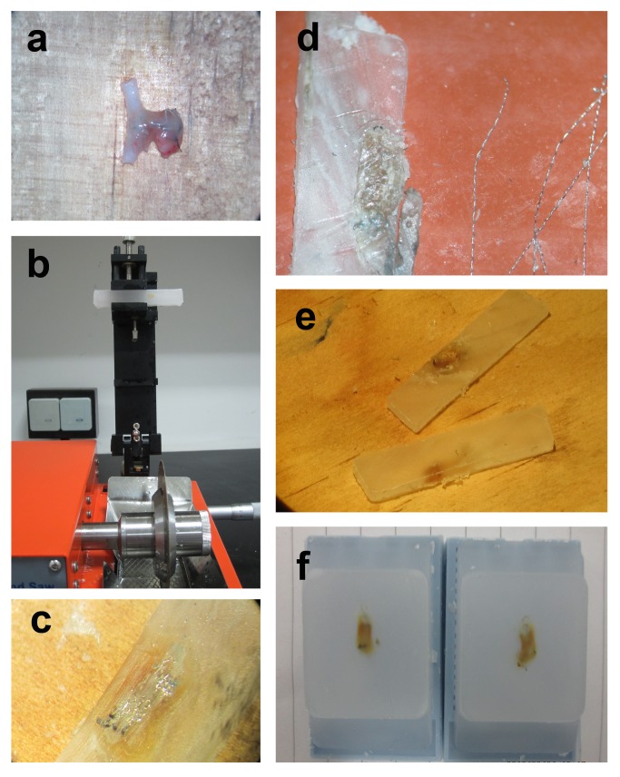
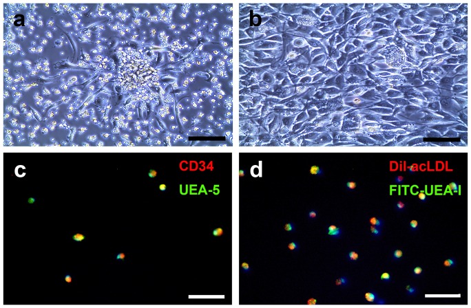
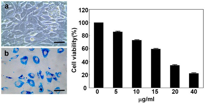
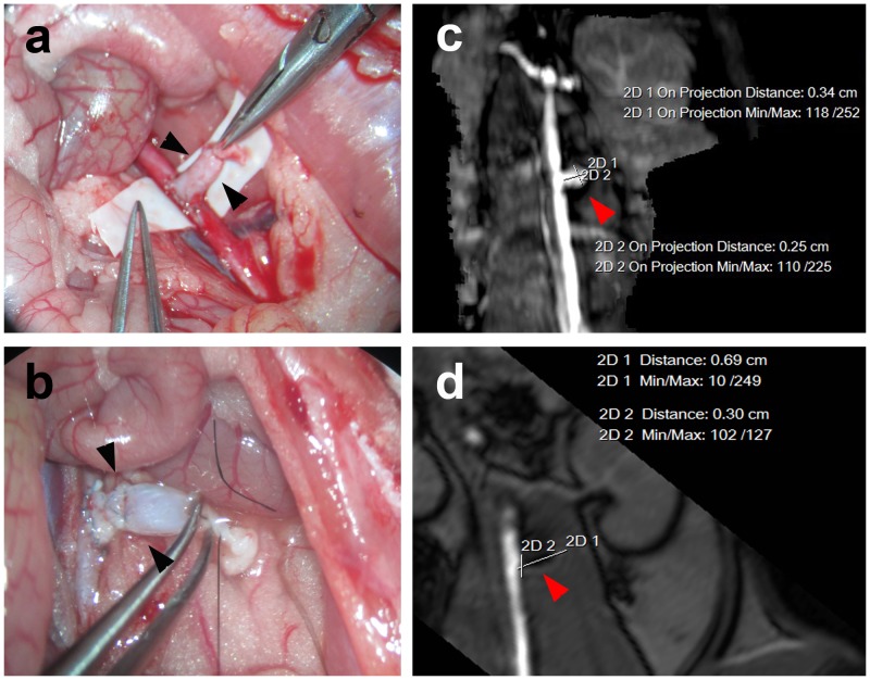
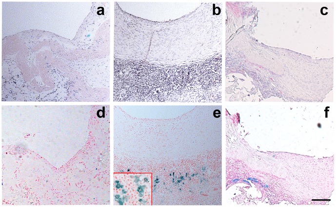
Aneurysm embolization with coil is now widely used clinically. However, the recurrence of aneurysms after embolization has always plagued neurosurgeons because the endothelial layer of the aneurysm neck loses its integrity after being embolized by coil. Bone marrow-derived endothelial progenitor cells (BM-EPCs) could be incorporated into injured endothelium and differentiate into mature endothelial cells during vascular repairing processes. The aim of our study is to explore the effects of BM-EPCs on aneurysm repairing and remodeling in a rat embolization model of abdominal aortic aneurysm. BM-EPC proliferation, migration and tube formation were not affected by super-paramagnetic iron oxide nanoparticle (SPIO) labeling compared to the controls (p>0.05). The number of SPIO-labeled cells greatly increased in EPC transplanted rats compared to that of phosphate buffered saline treated rats. SPIO-labeled EPC (SPIO-EPC) are mainly located in the aneurysm neck and surrounded by fibrous tissue. A histology study showed that the aneurysm orifice was closed with neointima and the aneurysm was filled with newly formed fibrous tissue. The SPIO-EPC accumulated in the aneurysm neck, which accelerated focal fibrous tissue remodeling, suggesting that BM-EPCs play a crucial role in repairing and remodeling the aneurysm neck orifice.
Conflict of interest statement
Figures
Similar articles
-
Erythropoietin Stimulates Endothelial Progenitor Cells to Induce Endothelialization in an Aneurysm Neck After Coil Embolization by Modulating Vascular Endothelial Growth Factor.Stem Cells Transl Med. 2016 Sep;5(9):1182-9. doi: 10.5966/sctm.2015-0264. Epub 2016 Jun 28. Stem Cells Transl Med. 2016. PMID: 27352930 Free PMC article.
-
Rosuvastatin for enhancement of aneurysm neck endothelialization after coil embolization: promotion of endothelial progenitor cells in a rodent model.J Neurosurg. 2016 May;124(5):1265-74. doi: 10.3171/2015.3.JNS142841. Epub 2015 Sep 25. J Neurosurg. 2016. PMID: 26406802
-
Superparamagnetic iron oxide nanoparticles may affect endothelial progenitor cell migration ability and adhesion capacity.Cytotherapy. 2010 Apr;12(2):251-9. doi: 10.3109/14653240903446910. Cytotherapy. 2010. PMID: 20196696
-
Effects of magnetically labeled exogenous endothelial progenitor cells on cerebral blood perfusion and microvasculature alterations after traumatic brain injury in rat model.Acta Radiol. 2013 Apr 1;54(3):313-23. doi: 10.1258/ar.2012.120605. Epub 2013 Mar 25. Acta Radiol. 2013. PMID: 23528570
-
Magnetic labeling, imaging and manipulation of endothelial progenitor cells using iron oxide nanoparticles.Future Med Chem. 2010 Mar;2(3):397-408. doi: 10.4155/fmc.09.165. Future Med Chem. 2010. PMID: 21426174 Review.
Cited by
-
Iron Oxide Nanoparticles in Regenerative Medicine and Tissue Engineering.Nanomaterials (Basel). 2021 Sep 8;11(9):2337. doi: 10.3390/nano11092337. Nanomaterials (Basel). 2021. PMID: 34578651 Free PMC article. Review.
-
Erythropoietin Stimulates Endothelial Progenitor Cells to Induce Endothelialization in an Aneurysm Neck After Coil Embolization by Modulating Vascular Endothelial Growth Factor.Stem Cells Transl Med. 2016 Sep;5(9):1182-9. doi: 10.5966/sctm.2015-0264. Epub 2016 Jun 28. Stem Cells Transl Med. 2016. PMID: 27352930 Free PMC article.
-
Preemptive Medicine for Cerebral Aneurysms.Neurol Med Chir (Tokyo). 2016 Sep 15;56(9):552-68. doi: 10.2176/nmc.st.2016-0063. Epub 2016 Apr 6. Neurol Med Chir (Tokyo). 2016. PMID: 27053328 Free PMC article. Review.
-
Histological and Transmission Electron Microscopy Results after Embolization with HydroSoft/HydroFrame Coils in Experimental Swine Aneurysm.Biomed Res Int. 2019 Dec 5;2019:4834535. doi: 10.1155/2019/4834535. eCollection 2019. Biomed Res Int. 2019. PMID: 31886218 Free PMC article.
-
Mechanisms of Healing in Coiled Intracranial Aneurysms: A Review of the Literature.AJNR Am J Neuroradiol. 2015 Jul;36(7):1216-22. doi: 10.3174/ajnr.A4175. Epub 2014 Nov 27. AJNR Am J Neuroradiol. 2015. PMID: 25430855 Free PMC article. Review.
References
-
- Juvela S (1992) Minor leak before rupture of an intracranial aneurysm and subarachnoid hemorrhage of unknown etiology. Neurosurgery 30: 7–11. - PubMed
-
- Fujinaka T, Yoshimine T, Mashimo T (2012) [Management of aneurysmal subarachnoid hemorrhage]. Masui 61: 962–970; discussion 970–962. - PubMed
-
- Aronson JP, Mitha AP, Hoh BL, Auluck PK, Pomerantseva I, et al. (2012) A novel tissue engineering approach using an endothelial progenitor cell-seeded biopolymer to treat intracranial saccular aneurysms. J Neurosurg 117: 546–554. - PubMed
-
- Kallmes DF, Helm GA, Hudson SB, Altes TA, Do HM, et al. (1999) Histologic evaluation of platinum coil embolization in an aneurysm model in rabbits. Radiology 213: 217–222. - PubMed
Publication types
MeSH terms
Substances
LinkOut - more resources
Full Text Sources
Other Literature Sources
Medical
