. 2014 Feb 28;9(2):e90085.
doi: 10.1371/journal.pone.0090085. eCollection 2014.

NAC, tiron and trolox impair survival of cell cultures containing glioblastoma tumorigenic initiating cells by inhibition of cell cycle progression

Affiliations

NAC, tiron and trolox impair survival of cell cultures containing glioblastoma tumorigenic initiating cells by inhibition of cell cycle progression

Massimiliano Monticone et al. PLoS One. .

Abstract

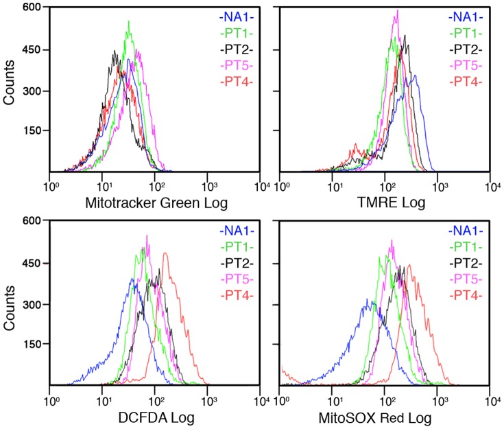
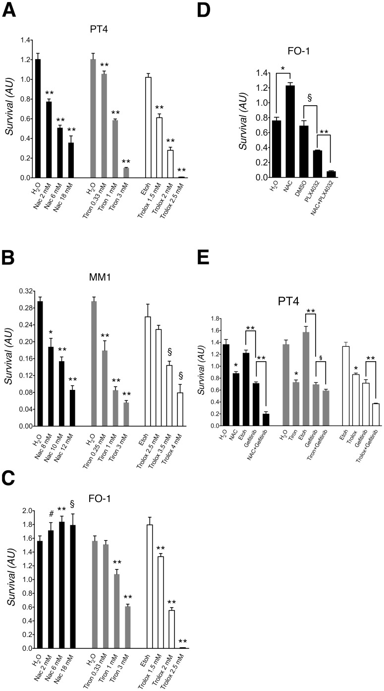
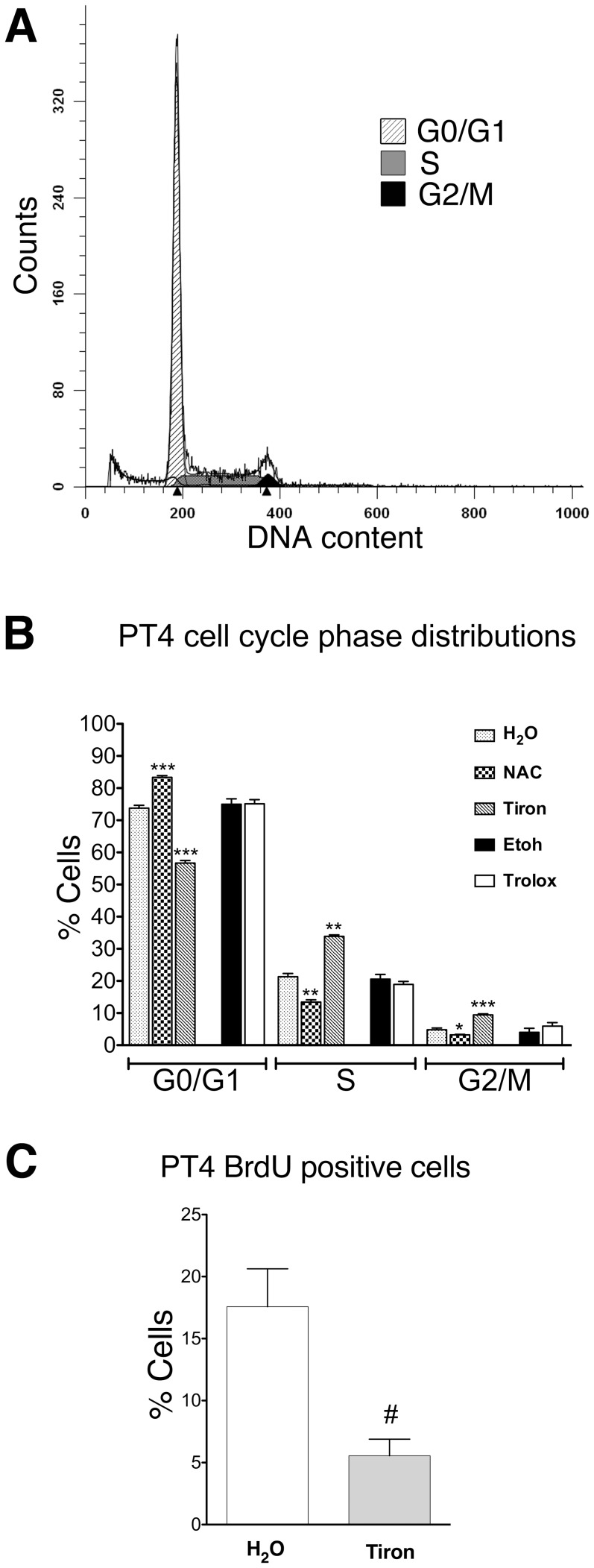
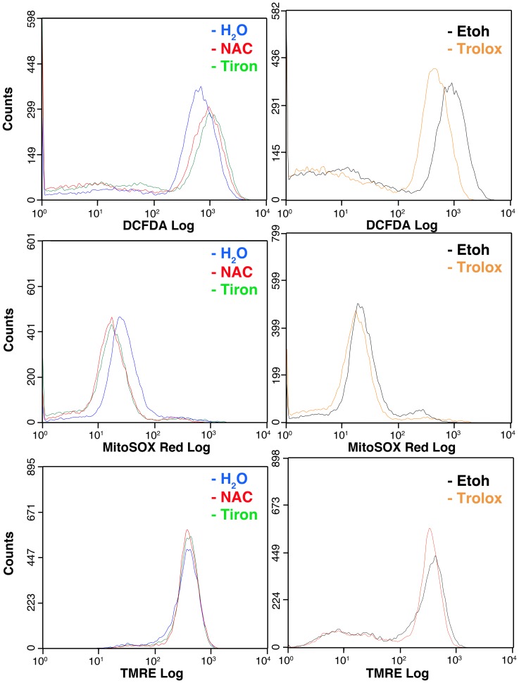
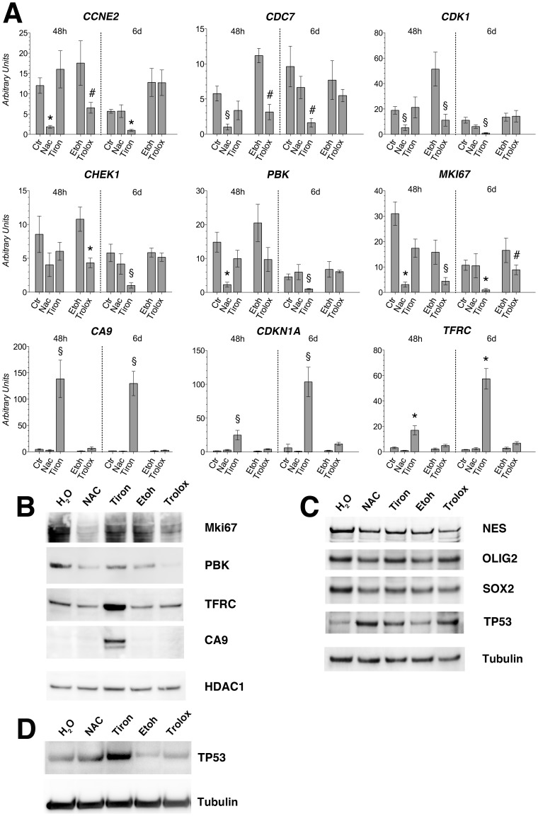
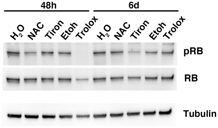
Reactive oxygen species (ROS) are metabolism by-products that may act as signaling molecules to sustain tumor growth. Antioxidants have been used to impair cancer cell survival. Our goal was to determine the mechanisms involved in the response to antioxidants of a human cell culture (PT4) containing glioblastoma (GBM) tumorigenic initiating cells (TICs). ROS production in the absence or presence of N-acetyl-L-cysteine (NAC), tiron, and trolox was evaluated by flow cytometry (FCM). The effects of these antioxidants on cell survival and apoptosis were evaluated by 3-(4,5-Dimethylthiazol-2-yl)-2,5-diphenyltetrazolium bromide assay (MTT) and FCM. The biological processes modulated by these drugs were determined by oligonucleotide microarray gene expression profiling. Our results showed that NAC, tiron and trolox impaired PT4 cell survival, had minor effects on ROS levels and caused wide deregulation of cell cycle genes. Furthermore, tiron and trolox caused inhibition of cell survival in two additional cell cultures containing TICs, FO-1 and MM1, established from a melanoma and a mesothelioma patient, respectively. NAC, instead, impaired survival of the MM1 cells but not of the FO-1 cells. However, when used in combination, NAC enhanced the inhibitory effect of PLX4032 (BRAF V600E inhibitor) and Gefitinib (EGFR inhibitor), on FO-1 and PT4 cell survival. Collectively, NAC, tiron and trolox modulated gene expression and impaired the growth of cultures containing TICs primarily by inhibiting cell cycle progression.


Conflict of interest statement

Competing Interests: The authors have declared that no competing interests exist.

Figures

Figure 1
Figure 1. Cell cultures containing GBM TICs display higher endogenous ROS generation than normal astrocytes.
Representative experiment of FCM analysis of normal human astrocytes (NA) and cell cultures containing GBM TICs obtained from four different patients (PT1, PT2, PT4, PT5) incubated with the indicated fluorescent probe. Mitotracker Green, TMRE, DCFDA and MitoSOX Red were used to evaluate mitochondrial mass, mitochondrial proton gradient, global ROS and mitochondrial superoxide, respectively.
Figure 2
Figure 2. Cell survival of the PT4, MM1 and FO-1 cell cultures containing TICs.
Survival of PT4 cells (A, E); MM1 cells (B) and FO-1 cells (C, D) after 6 days of treatment with the indicated substances and solvent controls. The medium renewal schedule was identical to that used for the cultures containing GBM TICs (see Introduction). Cell survival is expressed in arbitrary units as evaluated by MTT analysis. Standard deviations are indicated as vertical bars (n = 3 independent assays). DMSO concentration in (D) was 0.1% vol/vol. Drug concentrations in (D) were: NAC 20 mM, PLX4032 10 µM. Gefitinib final concentration in (E) was 3.9 µM. #The unpaired t-test was significant at P<0.05. §The unpaired t-test was significant at P = 0.01 or less. *The unpaired t-test was significant at P<0.001. **The unpaired t-test was significant at P<0.0001.
Figure 3
Figure 3. NAC and tiron modify the distribution of the cell cycle phases in the PT4 cell culture containing GBM TICs.
Representative experiment of high resolution FCM analysis of DAPI-stained nuclei of the H2O control PT4 culture containing GBM TICs after 48 h of exposure (A). Analysis of the percentage of PT4 cells in the cell cycle phases as determined by the ModFit LT™ software after 48 h of exposure with the indicated substances at the IC50 concentration and solvent controls (B). Percentage of PT4 cells in S phase actively synthesizing DNA after 48 h of exposure to tiron at the IC50 concentration with respect to control condition (H2O) performed by BrdU labeling and FCM analysis (C). Standard deviations are indicated as vertical bars (n = 4 independent assays, B ; n = 3 independent assays, C). #The unpaired t-test was significant at P<0.05. *The unpaired t-test was significant at P<0.001. **The unpaired t-test was significant at P<0.0001. ***The unpaired t-test was significant at P<0.00001.
Figure 4
Figure 4. NAC, tiron and trolox determine only modest changes in the ROS levels in the PT4 cell culture containing GBM TICs.
Representative experiment of FCM analysis of PT4 culture containing GBM TICs cells incubated with the indicated fluorescent probe after 48(containing NAC, tiron or trolox) replacement. DCFDA, MitoSOX Red and TMRE were used to evaluate global ROS, mitochondrial superoxide and mitochondrial proton gradient, respectively. This analysis showed that trolox reduced global cellular ROS levels but did not substantially modify mitochondrial superoxide levels. NAC and tiron, instead, slightly decreased mitochondrial superoxide levels while slightly enhancing global cellular ROS levels. This analysis also showed that the drugs used in this study did not induce changes of the mitochondrial proton gradient displayed by the PT4 cells in control conditions.
Figure 5
Figure 5. Validation of gene expression regulation by Real-time RT-PCR and Immunoblot analysis.
(A) Real-Time RT-PCR analysis performed with RNA extracted from the PT4 cell culture containing GBM TICs to validate the microarray data. This was accomplished on randomly selected genes from Table S3 and showed, in arbitrary units. Expression levels are relative to the expression of the housekeeping gene transcript coding for the ribosomal protein L19 (RPL19). Standard deviations are indicated as vertical bars (n = 3 independent assays). Gene name symbols are those approved by the Human Genome Organization Gene Nomenclature Committee (http://www.genenames.org/). Standard deviations are indicated as vertical bars (n = 3 independent assays). #The unpaired t-test was significant at P<0.05. §The unpaired t-test was significant at P<0.01. *The unpaired t-test was significant at P<0.001. (B) Western blot analyses were performed with lysates of the PT4 cell culture containing GBM TICs treated for 48 h with the indicated antioxidant drug and challenged with anti MKi67, Pdz-binding kinase (PBK), transferrin receptor (TFRC), carbonic anhydrase 9 (CA9). (C) Western blot analyses were performed with lysates of the PT4 cell culture containing GBM TICs treated for 6 days with the indicated antioxidant drug and challenged with anti nestin (NES), oligodendrocyte transcription factor 2 (OLIG2), SRY (sex determining region Y)-box 2 (SOX2) and tumor protein 53 (TP53) antibodies. (D) Western blot analyses were performed with lysates of the PT4 cell culture containing GBM TICs treated for 48 h with the indicated antioxidant drug and challenged with anti tumor protein 53 (TP53) mAb. (B–D) Immunoblotted membranes were subjected to multiple antibody challenging, stripping, control of effective stripping, and re-challenging with a different antibody. The last antibody used was an anti histone deacetylase 1 (HDAC1) (B) or an anti tubulin alpha (D) to show equal loading.
Figure 6
Figure 6. NAC, tiron and trolox treatments of the PT4 cultures containing GBM TICs inhibit the phosphorylation of the retinoblastoma (RB) protein.
Western blot analyses were performed with lysates of the PT4 cell culture containing GBM TICs treated for 48 h and 6 days with the indicated antioxidant drug. The membrane was challenged first with an anti phosphorylated RB (Ser 807/811) antibody, then with an anti total RB antibody and lastly with an anti tubulin antibody. Stripping and control of effective stripping were performed before re-challenging of the membrane.

References

    1. Freeman BA, Crapo JD (1982) Biology of disease: free radicals and tissue injury. Lab Invest 47: 412–426. - PubMed
    1. Jezek P, Hlavata L (2005) Mitochondria in homeostasis of reactive oxygen species in cell, tissues, and organism. Int J Biochem Cell Biol 37: 2478–2503. - PubMed
    1. Trachootham D, Lu W, Ogasawara MA, Nilsa RD, Huang P (2008) Redox regulation of cell survival. Antioxid Redox Signal 10: 1343–1374. - PMC - PubMed
    1. Bae YS, Oh H, Rhee SG, Yoo YD (2011) Regulation of reactive oxygen species generation in cell signaling. Mol Cells 32: 491–509. - PMC - PubMed
    1. Cosentino-Gomes D, Rocco-Machado N, Meyer-Fernandes JR (2012) Cell Signaling through Protein Kinase C Oxidation and Activation. Int J Mol Sci 13: 10697–10721. - PMC - PubMed

Publication types

MeSH terms

Substances