

Abrogation of TNFα production during cancer immunotherapy is crucial for suppressing side effects due to the systemic expression of IL-12
- PMID: 24587231
- PMCID: PMC3938584
- DOI: 10.1371/journal.pone.0090116
Abrogation of TNFα production during cancer immunotherapy is crucial for suppressing side effects due to the systemic expression of IL-12
Abstract
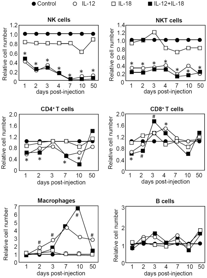
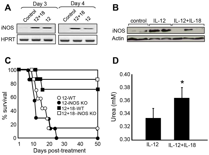
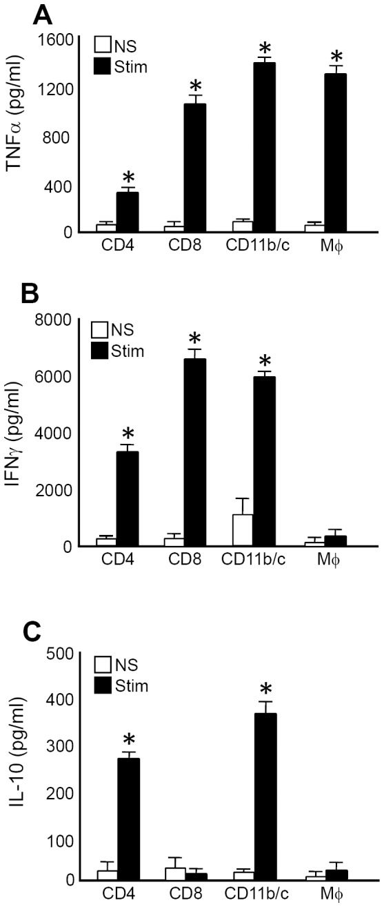
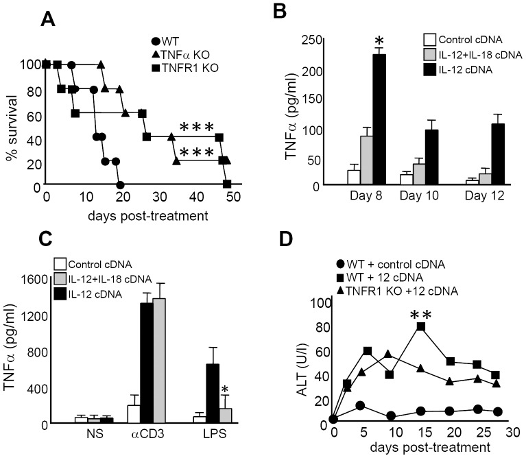
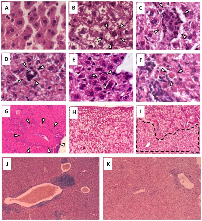
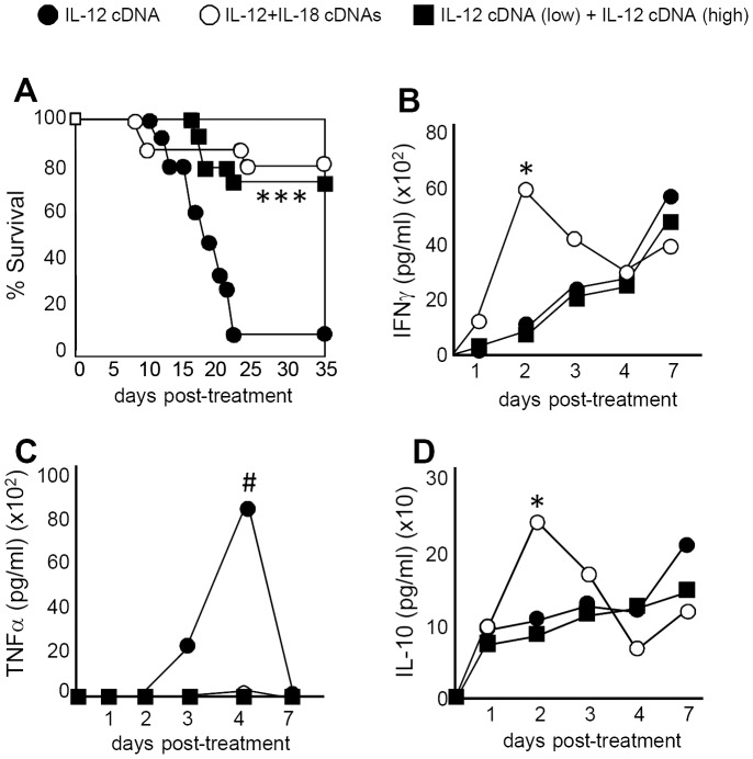
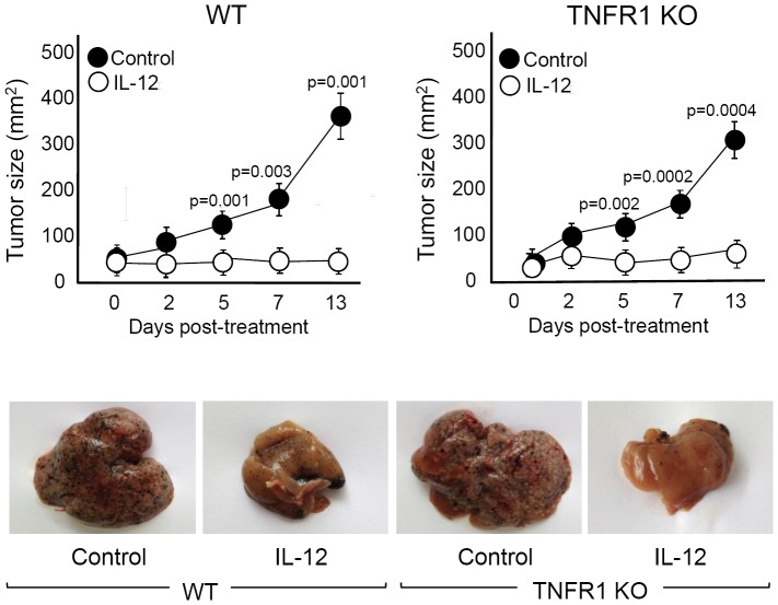
For more than a decade, the cytokine interleukin-12 (IL-12) has been utilized, either alone or in combination with other drugs, as a treatment for cancer. The numerous anti-tumor properties of IL-12 still generate interest in the clinical use of this cytokine, even though it has demonstrated toxicity when administrated systemically. As an approach to overcome this toxicity, numerous laboratories have attempted to induce IL-12 expression at the site of the tumor. However for tumors that are difficult to remove surgically or for the treatment of disseminated metastases, systemic expression of this cytokine still remains as the most efficient method of administration. Nevertheless, finding alternative approaches for the use of IL-12 in the treatment of cancer and unraveling the basis of IL-12-side effects remain a challenge. In the present work we demonstrate that systemic expression of IL-12 through hydrodynamic injection of IL-12 cDNA is able to induce different types of liver lesions associated with a toxic pathology. However we report here that hepatic toxicity is diminished and survival of mice enhanced in the absence of tumor necrosis factor alpha (TNFα). This observation is in contrast to several murine models and clinical trials that postulate interferon gamma (IFNγ) as the main cytokine responsible for IL-12 toxicity. Moreover, our work demonstrates that when IL-12 cDNA is co-injected with IL-18 cDNA or when mice are pre-treated with a low dose of IL-12 cDNA prior to receiving a high dose of IL-12 cDNA, systemic levels of TNFα are almost completely abrogated, resulting in improved survival and less hepatic damage. Importantly, abrogation of TNFα signaling does not affect the strong anti-tumor activity of IL-12. Thus, neutralizing TNFα with antagonists already approved for human use offers a promising approach to abrogate IL-12 side effects during the use of this cytokine for the treatment of cancer.
Conflict of interest statement
Figures
Similar articles
-
Coexpression of IL-18 strongly attenuates IL-12-induced systemic toxicity through a rapid induction of IL-10 without affecting its antitumor capacity.J Immunol. 2009 Jul 1;183(1):740-8. doi: 10.4049/jimmunol.0804166. Epub 2009 Jun 17. J Immunol. 2009. PMID: 19535628 Free PMC article.
-
IL-12 cDNA direct injection: antimetastatic effect from a single injection in a murine hepatic metastases model.J Surg Res. 2004 Dec;122(2):210-7. doi: 10.1016/j.jss.2004.04.021. J Surg Res. 2004. PMID: 15555620
-
Injection of complementary DNA encoding interleukin-12 inhibits tumor establishment at a distant site in a murine renal carcinoma model.Cancer Res. 1996 Aug 1;56(15):3399-403. Cancer Res. 1996. PMID: 8758901
-
Review: novel nonviral delivery approaches for interleukin-12 protein and gene systems: curbing toxicity and enhancing adjuvant activity.J Interferon Cytokine Res. 2006 Sep;26(9):593-608. doi: 10.1089/jir.2006.26.593. J Interferon Cytokine Res. 2006. PMID: 16978064 Review.
-
Interleukin-12 as an in situ cancer vaccine component: a review.Cancer Immunol Immunother. 2022 Sep;71(9):2057-2065. doi: 10.1007/s00262-022-03144-1. Epub 2022 Jan 13. Cancer Immunol Immunother. 2022. PMID: 35024897 Free PMC article. Review.
Cited by
-
Intra-tumoral production of IL18, but not IL12, by TCR-engineered T cells is non-toxic and counteracts immune evasion of solid tumors.Oncoimmunology. 2017 Oct 11;7(1):e1378842. doi: 10.1080/2162402X.2017.1378842. eCollection 2017. Oncoimmunology. 2017. PMID: 29296541 Free PMC article.
-
Safety levels of systemic IL-12 induced by cDNA expression as a cancer therapeutic.Immunotherapy. 2022 Feb;14(2):115-133. doi: 10.2217/imt-2021-0080. Epub 2021 Nov 16. Immunotherapy. 2022. PMID: 34783257 Free PMC article.
-
Preclinical validation: LV/IL-12 transduction of patient leukemia cells for immunotherapy of AML.Mol Ther Methods Clin Dev. 2016 Dec 7;3:16074. doi: 10.1038/mtm.2016.74. eCollection 2016. Mol Ther Methods Clin Dev. 2016. PMID: 27933304 Free PMC article.
-
Systemic sterile induced-co-expression of IL-12 and IL-18 drive IFN-γ-dependent activation of microglia and recruitment of MHC-II-expressing inflammatory monocytes into the brain.Int Immunopharmacol. 2022 Apr;105:108546. doi: 10.1016/j.intimp.2022.108546. Epub 2022 Jan 21. Int Immunopharmacol. 2022. PMID: 35074570 Free PMC article.
-
Interleukin-12 inhibits the hepatocellular carcinoma growth by inducing macrophage polarization to the M1-like phenotype through downregulation of Stat-3.Mol Cell Biochem. 2016 Apr;415(1-2):157-68. doi: 10.1007/s11010-016-2687-0. Epub 2016 Mar 22. Mol Cell Biochem. 2016. PMID: 27003285
References
-
- Rook AH, Zaki MH, Wysocka M, Wood GS, Duvic M, et al.. (2001) The role for interleukin-12 therapy of cutaneous T cell lymphoma. Ann N Y Acad Sci 941: : 177–184. PubMed: 11594571. - PubMed
-
- Hamid O, Solomon JC, Scotland R, Garcia M, Sian S, et al.. (2007) Alum with interleukin-12 augments immunity to a melanoma peptide vaccine: correlation with time to relapse in patients with resected high-risk disease. Clin Cancer Res 13: : 215–222. PubMed: 17200357. - PubMed
-
- Lenzi R, Edwards R, June C, Seiden MV, Garcia ME, et al.. (2007) Phase II study of intraperitoneal recombinant interleukin-12 (rhIL-12) in patients with peritoneal carcinomatosis (residual disease < 1 cm) associated with ovarian cancer or primary peritoneal carcinoma.J Transl Med 5: : 66. doi: 10.1186/1479-5876-5-66. PM:18076766. - PMC - PubMed
-
- Yarchoan R, Pluda JM, Wyvill KM, Aleman K, Rodriguez-Chavez IR, et al.. (2007) Treatment of AIDS-related Kaposi's sarcoma with interleukin-12: rationale and preliminary evidence of clinical activity. Crit Rev Immunol 27: : 401–414. PubMed: 18197804. - PubMed
Publication types
MeSH terms
Substances
Grants and funding
LinkOut - more resources
Full Text Sources
Other Literature Sources
Molecular Biology Databases
Research Materials
Miscellaneous
