. 2014 Feb 25;9(2):e90141.
doi: 10.1371/journal.pone.0090141. eCollection 2014.

Stathmin protein level, a potential predictive marker for taxane treatment response in endometrial cancer

Affiliations

Stathmin protein level, a potential predictive marker for taxane treatment response in endometrial cancer

Henrica M J Werner et al. PLoS One. .

Abstract

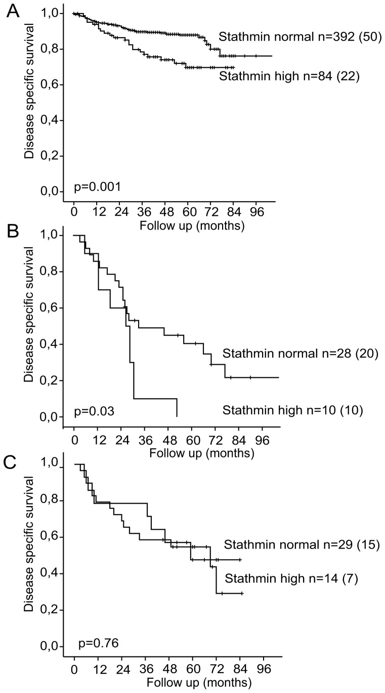
Stathmin is a prognostic marker in many cancers, including endometrial cancer. Preclinical studies, predominantly in breast cancer, have suggested that stathmin may additionally be a predictive marker for response to paclitaxel. We first evaluated the response to paclitaxel in endometrial cancer cell lines before and after stathmin knock-down. Subsequently we investigated the clinical response to paclitaxel containing chemotherapy in metastatic endometrial cancer in relation to stathmin protein level in tumors. Stathmin level was also determined in metastatic lesions, analyzing changes in biomarker status on disease progression. Knock-down of stathmin improved sensitivity to paclitaxel in endometrial carcinoma cell lines with both naturally higher and lower sensitivity to paclitaxel. In clinical samples, high stathmin level was demonstrated to be associated with poor response to paclitaxel containing chemotherapy and to reduced disease specific survival only in patients treated with such combination. Stathmin level increased significantly from primary to metastatic lesions. This study suggests, supported by both preclinical and clinical data, that stathmin could be a predictive biomarker for response to paclitaxel treatment in endometrial cancer. Re-assessment of stathmin level in metastatic lesions prior to treatment start may be relevant. Also, validation in a randomized clinical trial will be important.


Conflict of interest statement

Competing Interests: The authors report the following conflicts: The University of Bergen/Haukeland University Hospital/Dana Farber Cancer Institute/Harvard through BTO (Bergen Teknologioverføring AS): Some aspects relating to STMN1 as prognostic and predictive marker for endometrial cancer are subject to pending intellectual property rights (US 12/962,946). This does not alter the authors' adherence to the PLOS ONE policies on sharing data and materials.

Figures

Figure 1
Figure 1. Sensitivity of wild-type cell lines to paclitaxel treatment.
A: Microscopic assessment of apoptosis in Ishikawa and Hec1B wild-type cells after treatment with paclitaxel in the following dosages: 0 nM, 5 nM, 10 nM, 50 nM, 100 nM, 250 nM and 500 nM. Results are representative of 5 independent experiments. Standard errors of the mean are indicated. B: Cell metabolic activity assessed with a proliferation assay (MTS, Promega) in Ishikawa and Hec1B wild-type cells after treatment with paclitaxel in the following dosages: 0 nM, 1 nM, 10 nM, 50 nM, 100 nM, 250 nM and 500 nM. Results are representative of 3 (Ishikawa) and 2 (Hec1B) independent experiments.
Figure 2
Figure 2. Ishikawa cell line experiments after stathmin knock-down.
A: Immunoblot after transfecting cells with a stathmin lentiviral shRNAmir (‘stmn kd’) or a non-silencing control (‘non sil’) as well as the parental cell line (wild-type; ‘WT’). Blots were stained for stathmin, and β-actin for loading control. B: Ishikawa wild-type cell line, non-silencing and stathmin knock-down after treatment with paclitaxel for 24 h in the following dosages: 0 nM, 5 nM, 10 nM, 50 nM, 100 nM, 250 nM and 500 nM. The level of fragmentation of the cells is indicated in an insert, as a proxy of progression in the apoptotic process. Diamonds; wild-type, triangles; non-silencing and crosses; stathmin knock-down cells. C: Immunoblot of Ishikawa wild-type, control (non-silencing) and stathmin knock-down cell lines after treatment with paclitaxel for 24 h in the following dosages: 0 nM, 100 nM and 250 nM. The blot was stained for cleaved PARP and stathmin, with β-actin serving as loading control. D: Left: Ishikawa wild-type (‘WT’) cell line and Right: Ishikawa stathmin knock-down (‘Stmn kd’) cell line. Microscopic images of cells after treatment for 24 h with 0 nM (top row) or 500 nM (bottom row) paclitaxel also demonstrating increased fragmentation rate for the stathmin knock-down Ishikawa cells (right lower panel) compared to wild-type (left lower panel).
Figure 3
Figure 3. Hec1B cell line experiments after stathmin knock-down.
A: Immunoblot after transfecting cells with a stathmin lentiviral shRNAmir (‘stmn kd’) or a non-silencing control (‘non sil’) as well as the parental cell line (wild-type; ‘WT’). Blots were stained for stathmin, and β-actin for loading control. B: Hec1B wild-type cell line, non-silencing and stathmin knock-down lines, after treatment with paclitaxel for 24 h in the following dosages: 0 nM, 5 nM, 10 nM, 50 nM, 100 nM, 250 nM and 500 nM. C: Left: Hec1B wild-type (‘WT’) cell line and Right: Hec1B stathmin knock-down (‘Stmn kd’) cell line. Showing microscopic images of cells after treatment for 24 h with 0 nM (top row) or 500 nM (bottom row) paclitaxel demonstrating increased cell death for the stathmin knock-down Hec1B cells (right lower panel) compared to Hec1B wild-type (left lower panel).
Figure 4
Figure 4. Stathmin protein expression in relation to clinical parameters.
A: Pictures representative of weak immunohistochemical stathmin staining (top) and strong (high or pathologic) stathmin staining (bottom) in endometrial carcinoma. Bars (right lower corner) measure 40 μm. B: Clinical response to paclitaxel for all endometrial carcinoma patients with evaluable disease according to RECIST criteria and separated for normal versus high stathmin level. Poor response (RECIST: static disease or disease progression) indicated in blue, good response (RECIST: complete or partial response) indicated in red. C: Comparison of high (pathologic) stathmin protein level in primary and metastatic lesions.
Figure 5
Figure 5. Disease specific survival after primary treatment for endometrial carcinoma patients (Kaplan-Meier curves) related to stathmin protein expression by IHC in primary tumor.
A: All patients with complete data (n = 476). Number of disease specific events between brackets. B: All patients with metastatic disease who received paclitaxel treatment (n = 38). Number of disease specific events between brackets. C: All patients with metastatic disease who received different treatments (n = 43). Number of disease specific events between brackets.

Similar articles

Cited by

References

    1. Belletti B, Baldassarre G (2011) Stathmin: a protein with many tasks. New biomarker and potential target in cancer. Expert opinion on therapeutic targets 15: 1249–1266. - PubMed
    1. Marklund U, Larsson N, Gradin HM, Brattsand G, Gullberg M (1996) Oncoprotein 18 is a phosphorylation-responsive regulator of microtubule dynamics. The EMBO journal 15: 5290–5298. - PMC - PubMed
    1. Mistry SJ, Li HC, Atweh GF (1998) Role for protein phosphatases in the cell-cycle-regulated phosphorylation of stathmin. The Biochemical journal 334 (Pt 1): 23–29. - PMC - PubMed
    1. Rubin CI, Atweh GF (2004) The role of stathmin in the regulation of the cell cycle. Journal of cellular biochemistry 93: 242–250. - PubMed
    1. Biomarkers Definitions Working G (2001) Biomarkers and surrogate endpoints: preferred definitions and conceptual framework. Clinical pharmacology and therapeutics 69: 89–95. - PubMed

Publication types

MeSH terms

LinkOut - more resources