Comparative Study
. 2014 Feb 28;9(2):e90398.
doi: 10.1371/journal.pone.0090398. eCollection 2014.

Human and mouse skeletal muscle stem cells: convergent and divergent mechanisms of myogenesis

Affiliations
Comparative Study

Human and mouse skeletal muscle stem cells: convergent and divergent mechanisms of myogenesis

Akshay Bareja et al. PLoS One. .

Abstract

Satellite cells are the chief contributor to skeletal muscle growth and regeneration. The study of mouse satellite cells has accelerated in recent years due to technical advancements in the isolation of these cells. The study of human satellite cells has lagged and thus little is known about how the biology of mouse and human satellite cells compare. We developed a flow cytometry-based method to prospectively isolate human skeletal muscle progenitors from the satellite cell pool using positive and negative selection markers. Results show that this pool is enriched in PAX7 expressing cells that possess robust myogenic potential including the ability to give rise to de novo muscle in vivo. We compared mouse and human satellite cells in culture and identify differences in the elaboration of the myogenic genetic program and in the sensitivity of the cells to cytokine stimulation. These results indicate that not all mechanisms regulating mouse satellite cell activation are conserved in human satellite cells and that such differences may impact the clinical translation of therapeutics validated in mouse models. Thus, the findings of this study are relevant to developing therapies to combat muscle disease.


Conflict of interest statement

Competing Interests: The authors have the following conflicts: JAH, GL, WJE, JMF and ANB are employees of GlaxoSmithKline (GSK). CC, JL, and ACH are or were employees of Five Prime Therapeutics (FP). AB was a Duke University postdoctoral fellow funded by GlaxoSmithKline. Funding for this research by a commercial source does not alter the authors' adherence to all the PLOS ONE policies on sharing data and materials.

Figures

Figure 1
Figure 1. huSMP isolation strategy.
A. FACS plot of Calcein blue (live cells) and Propidium Iodide (dead cells) indicating events gated as live cells. B. FACS plot of cells before depletion of the FITC-linked antibody labeled cells. C. FACS plot demonstrating the effectiveness of FITC+ linked antibody labeled cell depletion. D. FACS plot of a sample depleted of FITC+ labeled cells gated on CD56+ cells. E. FACS plot of the CD56+ subset from D showing the CXCR4+, CD56+ subset of cells that were collected for further experimentation.
Figure 2
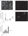
Figure 2. Myogenic activity of transplanted cells.
A. Myotube formation by huSMPs after 4 days of differentiation indicated by myosin heavy chain staining. Scale bar = 100 µm. B. qPCR data of RNA extracted from transplanted mouse gastrocnemius muscle. A1, A5, L1, L8 represent individual mice transplanted with two different preparations of huSMPs into the gastrocnemius muscle of one leg. Expression of human myogenic genes is observed in three of the four samples. Relative RNA amount was obtained after normalization to mouse Gapdh levels. C. Mouse MyoD1 expression in all samples indicates that the RNA was of sufficient quality for qPCR analysis in all samples. D. No human DMD+ fibers were observed in control contralateral muscle injected with growth media only. E. Human DMD protein expression in mouse myofibers after transplantation of huSMPs indicates engraftment and differentiation of the cells (arrows). Scale bar = 50 µm.
Figure 3
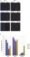
Figure 3. Temporal regulation of Myogenic transcription factor expression.
A. Immunofluorescence staining of cultured huSMPs shows increased markers of activation over time (scale bar = 50 µm). B. Quantification of positive staining nuclei as a percentage of total nuclei in the wells.
Figure 4
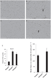
Figure 4. No evidence of lineage changes in huSMP cultures.
A. Phase contrast photomicrograph of a proliferating culture of huSMPs just prior to changing to differentiation conditions. B. After 7 days of differentiation in myogenic differentiation media myotubes are readily observed (arrow). C. In adipogenic differentiation media huSMPs do not fuse into myotubes and do not exhibit morphological characteristics of adipocytes such as intracellular lipid droplets. D. In osteogenic media huSMPs still differentiate into myotubes (arrow). E. The expression of MYOD1 RNA is maintained in all three differentiation conditions. F. Total area of myosin heavy chain staining normalized to the number of nuclei per well is not altered by differentiation in osteogenic media. Adipogenic media appears to block progression to myosin heavy chain expressing myoblast and myotubes. Scale bar = 200 µm.
Figure 5
Figure 5. Comparison of mouse and human SMP myogenic programs.
A. Gene expression data derived from qPCR assays run with RNA extracted from huSMPs at the indicated time points displayed as a heat map. Numerous differences in the timing of expression of key myogenic genes are readily observed. B. Mouse SMPs cultured in the presence of mouse IL1β (1 ng/ml) have 2.5 times as many SMPs at the end of a five day culture period as compared to cell cultured under standard conditions. Mouse SMPs lacking the IL1β receptor do not respond to the cytokine indicating specificity of the growth response. C. Growth of human SMPs derived from three different donors is reduced by culture in the presence of human IL1β (125 pg/ml).
Figure 6
Figure 6. Knockdown of myogenic transcription factors by siRNA reveals regulatory interactions.
A. The myogenic transcription factor analyzed by qPCR is listed on the left. siRNA treatments are on the X axis of each graph. Each symbol represents huSMPs derived from a single donor (a total of three donors tested). The matched-pair analysis clearly indicates the knockdown of the cognate transcripts (C, D, and E). B. A significant reduction in MYF5 RNA as a result of siMYOD1 treatment is observed. D. MYF5 expression is unchanged with siPAX7 a result that is in contrast to that observed in the mouse.

References

    1. Mauro A (1961) Satellite cell of skeletal muscle fibers. J Biophys Biochem Cytol 9: 493–495. - PMC - PubMed
    1. Boldrin L, Muntoni F, Morgan JE (2010) Are human and mouse satellite cells really the same? J Histochem Cytochem 58: 941–955. - PMC - PubMed
    1. Lepper C, Partridge TA, Fan CM (2011) An absolute requirement for Pax7-positive satellite cells in acute injury-induced skeletal muscle regeneration. Development 138: 3639–3646. - PMC - PubMed
    1. Sherwood RI, Christensen JL, Conboy IM, Conboy MJ, Rando TA, et al. (2004) Isolation of adult mouse myogenic progenitors: functional heterogeneity of cells within and engrafting skeletal muscle. Cell 119: 543–554. - PubMed
    1. Yi L, Rossi F (2011) Purification of progenitors from skeletal muscle. J Vis Exp 2476 [pii] 10.3791/2476. - PMC - PubMed

Publication types

MeSH terms