HIV-1 entry and trans-infection of astrocytes involves CD81 vesicles
- PMID: 24587404
- PMCID: PMC3938779
- DOI: 10.1371/journal.pone.0090620
HIV-1 entry and trans-infection of astrocytes involves CD81 vesicles
Abstract
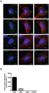
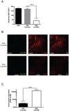
Astrocytes are extensively infected with HIV-1 in vivo and play a significant role in the development of HIV-1-associated neurocognitive disorders. Despite their extensive infection, little is known about how astrocytes become infected, since they lack cell surface CD4 expression. In the present study, we investigated the fate of HIV-1 upon infection of astrocytes. Astrocytes were found to bind and harbor virus followed by biphasic decay, with HIV-1 detectable out to 72 hours. HIV-1 was observed to associate with CD81-lined vesicle structures. shRNA silencing of CD81 resulted in less cell-associated virus but no loss of co-localization between HIV-1 and CD81. Astrocytes supported trans-infection of HIV-1 to T-cells without de novo virus production, and the virus-containing compartment required 37°C to form, and was trypsin-resistant. The CD81 compartment observed herein, has been shown in other cell types to be a relatively protective compartment. Within astrocytes, this compartment may be actively involved in virus entry and/or spread. The ability of astrocytes to transfer virus, without de novo viral synthesis suggests they are capable of sequestering and protecting virus and thus, they could potentially facilitate viral dissemination in the CNS.
Conflict of interest statement
Figures


Similar articles
-
A role for CD81 on the late steps of HIV-1 replication in a chronically infected T cell line.Retrovirology. 2009 Mar 11;6:28. doi: 10.1186/1742-4690-6-28. Retrovirology. 2009. PMID: 19284574 Free PMC article.
-
Tetraspanin CD81 regulates HSV-1 infection.Med Microbiol Immunol. 2020 Aug;209(4):489-498. doi: 10.1007/s00430-020-00684-0. Epub 2020 Jun 4. Med Microbiol Immunol. 2020. PMID: 32500359 Free PMC article.
-
CD81 large extracellular loop-containing fusion proteins with a dominant negative effect on HCV cell spread and replication.J Gen Virol. 2017 Jul;98(7):1646-1657. doi: 10.1099/jgv.0.000850. Epub 2017 Jul 19. J Gen Virol. 2017. PMID: 28721844
-
CD81-receptor associations--impact for hepatitis C virus entry and antiviral therapies.Viruses. 2014 Feb 18;6(2):875-92. doi: 10.3390/v6020875. Viruses. 2014. PMID: 24553110 Free PMC article. Review.
-
CD81 and hepatitis C virus (HCV) infection.Viruses. 2014 Feb 6;6(2):535-72. doi: 10.3390/v6020535. Viruses. 2014. PMID: 24509809 Free PMC article. Review.
Cited by
-
The Potential of the CNS as a Reservoir for HIV-1 Infection: Implications for HIV Eradication.Curr HIV/AIDS Rep. 2015 Jun;12(2):299-303. doi: 10.1007/s11904-015-0257-9. Curr HIV/AIDS Rep. 2015. PMID: 25869939 Review.
-
Potential pharmacological approaches for the treatment of HIV-1 associated neurocognitive disorders.Fluids Barriers CNS. 2020 Jul 10;17(1):42. doi: 10.1186/s12987-020-00204-5. Fluids Barriers CNS. 2020. PMID: 32650790 Free PMC article. Review.
-
General Pathophysiology of Astroglia.Adv Exp Med Biol. 2019;1175:149-179. doi: 10.1007/978-981-13-9913-8_7. Adv Exp Med Biol. 2019. PMID: 31583588 Free PMC article. Review.
-
Astrocytes in human central nervous system diseases: a frontier for new therapies.Signal Transduct Target Ther. 2023 Oct 13;8(1):396. doi: 10.1038/s41392-023-01628-9. Signal Transduct Target Ther. 2023. PMID: 37828019 Free PMC article. Review.
-
Morphine exposure exacerbates HIV-1 Tat driven changes to neuroinflammatory factors in cultured astrocytes.PLoS One. 2020 Mar 25;15(3):e0230563. doi: 10.1371/journal.pone.0230563. eCollection 2020. PLoS One. 2020. PMID: 32210470 Free PMC article.
References
-
- Gonzalez-Scarano F, Martin-Garcia J (2005) The neuropathogenesis of AIDS. Nat Rev Immunol 5: 69–81. - PubMed
-
- Brew BJ, Gray L, Lewin S, Churchill M (2013) Is specific HIV eradication from the brain possible or needed? Expert Opin Biol Ther 13: 403–409. - PubMed
-
- Takahashi K, Wesselingh SL, Griffin DE, McArthur JC, Johnson RT, et al. (1996) Localization of HIV-1 in human brain using polymerase chain reaction/in situ hybridization and immunocytochemistry. Ann Neurol 39: 705–711. - PubMed
Publication types
MeSH terms
Substances
LinkOut - more resources
Full Text Sources
Other Literature Sources
Research Materials

