Conservative management of idiopathic anterior atlantoaxial subluxation without neurological deficits in an 83-year-old female: A case report
- PMID: 24587500
- PMCID: PMC3924511
Conservative management of idiopathic anterior atlantoaxial subluxation without neurological deficits in an 83-year-old female: A case report
Abstract
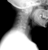
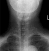
Atlantoaxial subluxation that is not related to traumatic, congenital, or rheumatological conditions is rare and can be a diagnostic challenge. This case report details a case of anterior atlantoaxial subluxation in an 83-year-old female without history of trauma, congenital, or rheumatological conditions. She presented to the chiropractor with insidious neck pain and headaches, without neurological deficits. Radiographs revealed a widened atlantodental space (measuring 6 mm) indicating anterior atlantoaxial subluxation and potential sagittal atlantoaxial instability. Prompt detection and appropriate conservative management resulted in favourable long-term outcome at 13-months follow-up. Conservative management included education, mobilizations, soft tissue therapy, monitoring for neurological progression, and co-management with the family physician. The purpose of this case report is to heighten awareness of the clinical presentation of idiopathic anterior atlantoaxial subluxation without neurological deficits. Discussion will focus on the incidence, mechanism, clinical presentation, and conservative management of a complex case of anterior atlantoaxial subluxation.
La subluxation atloïdo axoïdienne qui n’est pas liée à des conditions traumatiques, congénitales ou rhumatologiques est rare et peut présenter un défi sur le plan du diagnostic. Cette étude de cas décrit un cas de subluxation atloïdo axoïdienne antérieure chez une femme de 83 ans sans antécédents de pathologies traumatiques, congénitales ou rhumatismales. Elle s’est présentée chez le chiropraticien avec des douleurs cervicales insidieuses et des maux de tête, sans déficits neurologiques. Les radiographies ont révélé un espace atlanto-dental élargi (6 mm) indiquant une subluxation atloïdo axoïdienne antérieure et la possibilité d’une instabilité atloïdo axoïdienne sagittale. La détection rapide et un traitement conservateur approprié ont mené à un résultat favorable à long terme, avec un suivi après 13 mois. Le traitement conservateur comprend la sensibilisation, les mobilisations, le traitement des tissus mous, le suivi de la progression neurologique, et la cogestion avec le médecin de famille. Cette étude de cas vise à la sensibilisation de la présentation clinique d’une subluxation atloïdo axoïdienne antérieure idiopathique sans déficits neurologiques. La discussion portera sur l’incidence, le mécanisme, la présentation clinique et le traitement conservateur d’un cas complexe de subluxation atloïdo axoïdienne antérieure.
Keywords: atlantoaxial subluxation; atraumatic; conservative management; idiopathic; upper cervical.
Figures


Similar articles
-
Group A Streptococcus dysgalactiae subspecies equisimilis vertebral osteomyelitis accompanied by progressive atlantoaxial subluxation: A case report and literature review.Medicine (Baltimore). 2023 Aug 25;102(34):e34968. doi: 10.1097/MD.0000000000034968. Medicine (Baltimore). 2023. PMID: 37653834 Free PMC article. Review.
-
Congenital anterior midline cleft of the atlas and posterior atlanto-occipital fusion associated with symptomatic anterior atlantoaxial subluxation.Eur J Orthop Surg Traumatol. 2012 Nov;22 Suppl 1:35-9. doi: 10.1007/s00590-012-1011-2. Epub 2012 May 25. Eur J Orthop Surg Traumatol. 2012. PMID: 26662745
-
Spontaneous atlantoaxial subluxation associated with tonsillitis.Asian J Neurosurg. 2015 Apr-Jun;10(2):139-41. doi: 10.4103/1793-5482.152112. Asian J Neurosurg. 2015. PMID: 25972950 Free PMC article.
-
Atraumatic atlantoaxial subluxation in pediatric enthesitis-related juvenile idiopathic arthritis: illustrative case.J Neurosurg Case Lessons. 2025 May 19;9(20):CASE25121. doi: 10.3171/CASE25121. Print 2025 May 19. J Neurosurg Case Lessons. 2025. PMID: 40388885 Free PMC article.
-
Traumatic anterior atlantoaxial subluxation occurring in a professional rugby athlete: case report and review of literature related to atlantoaxial injuries in sports activities.Spine (Phila Pa 1976). 2004 Feb 1;29(3):E61-4. doi: 10.1097/01.brs.0000106498.80385.9a. Spine (Phila Pa 1976). 2004. PMID: 14752366 Review.
Cited by
-
Effects of Two Exercise Regimes on Patients with Chiari Malformation Type 1: a Randomized Controlled Trial.Cerebellum. 2023 Apr;22(2):305-315. doi: 10.1007/s12311-022-01397-1. Epub 2022 Mar 24. Cerebellum. 2023. PMID: 35325392 Clinical Trial.
-
Atlantoaxial Rotatory Subluxation in a 10-Year-Old Boy.Clin Med Insights Arthritis Musculoskelet Disord. 2020 Jul 1;13:1179544120939069. doi: 10.1177/1179544120939069. eCollection 2020. Clin Med Insights Arthritis Musculoskelet Disord. 2020. PMID: 32655279 Free PMC article.
-
Secondary atlantoaxial subluxation in isolated cervical dystonia-a case report.AME Case Rep. 2020 Apr 30;4:9. doi: 10.21037/acr.2020.03.03. eCollection 2020. AME Case Rep. 2020. PMID: 32420532 Free PMC article.
-
Atlantoaxial Subluxation Related to Axial Spondylarthritis: A Case-Based Systematic Review.Mediterr J Rheumatol. 2024 Dec 31;35(4):563-572. doi: 10.31138/mjr.070624.asr. eCollection 2024 Dec. Mediterr J Rheumatol. 2024. PMID: 39886282 Free PMC article. Review.
-
Multimodal Management of Coexisting Atlantoaxial Subluxation and Spinal Stenosis in an Older Adult: A Case Report and Literature Review.Cureus. 2024 Jan 1;16(1):e51442. doi: 10.7759/cureus.51442. eCollection 2024 Jan. Cureus. 2024. PMID: 38298323 Free PMC article.
References
-
- Klimo P, Jr, Kan P, Rao G, Apfelbaum R, Brockmeyer D. Os odontoideum: presentation, diagnosis, and treatment in a series of 78 patients. J Neurosurg Spine. 2008;9:332–42. - PubMed
-
- Wasserman BR, Moskovich R, Razi AE. Rheumatoid arthritis of the cervical spine – clinical considerations. Bull NYU Hosp Jt Dis. 2011;69:136–48. - PubMed
-
- Nakajima K, Onomura T, Tanida Y, Ishibashi I. Factors related to the severity of myelopathy in atlantoaxial instability. Spine. 1996;21:1440–5. - PubMed
LinkOut - more resources
Full Text Sources
