

Multifunctional to multistage delivery systems: The evolution of nanoparticles for biomedical applications
- PMID: 24587616
- PMCID: PMC3938208
- DOI: 10.1007/s11434-012-5387-5
Multifunctional to multistage delivery systems: The evolution of nanoparticles for biomedical applications
Abstract
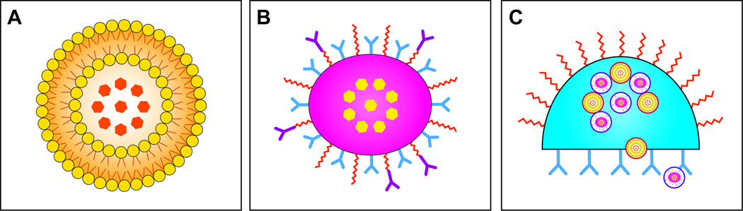
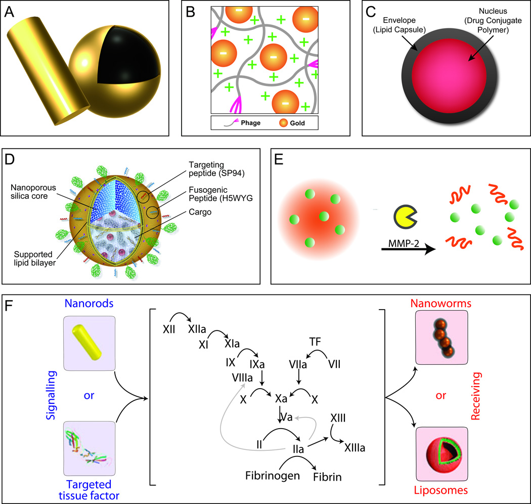
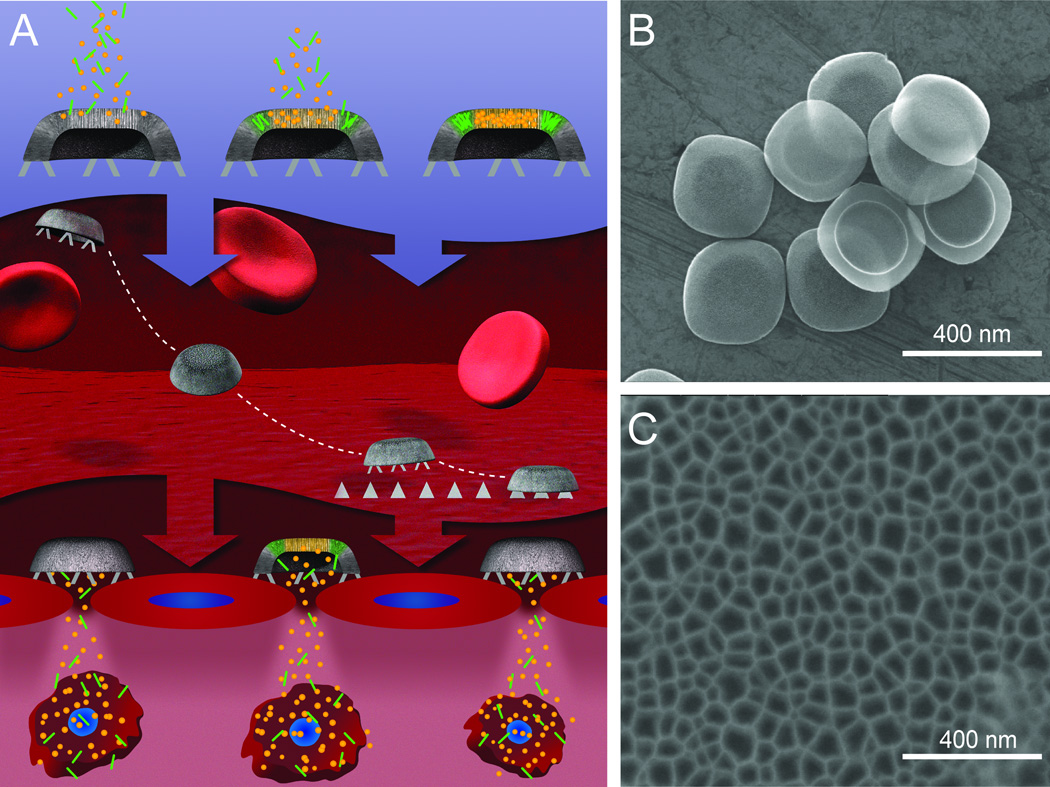
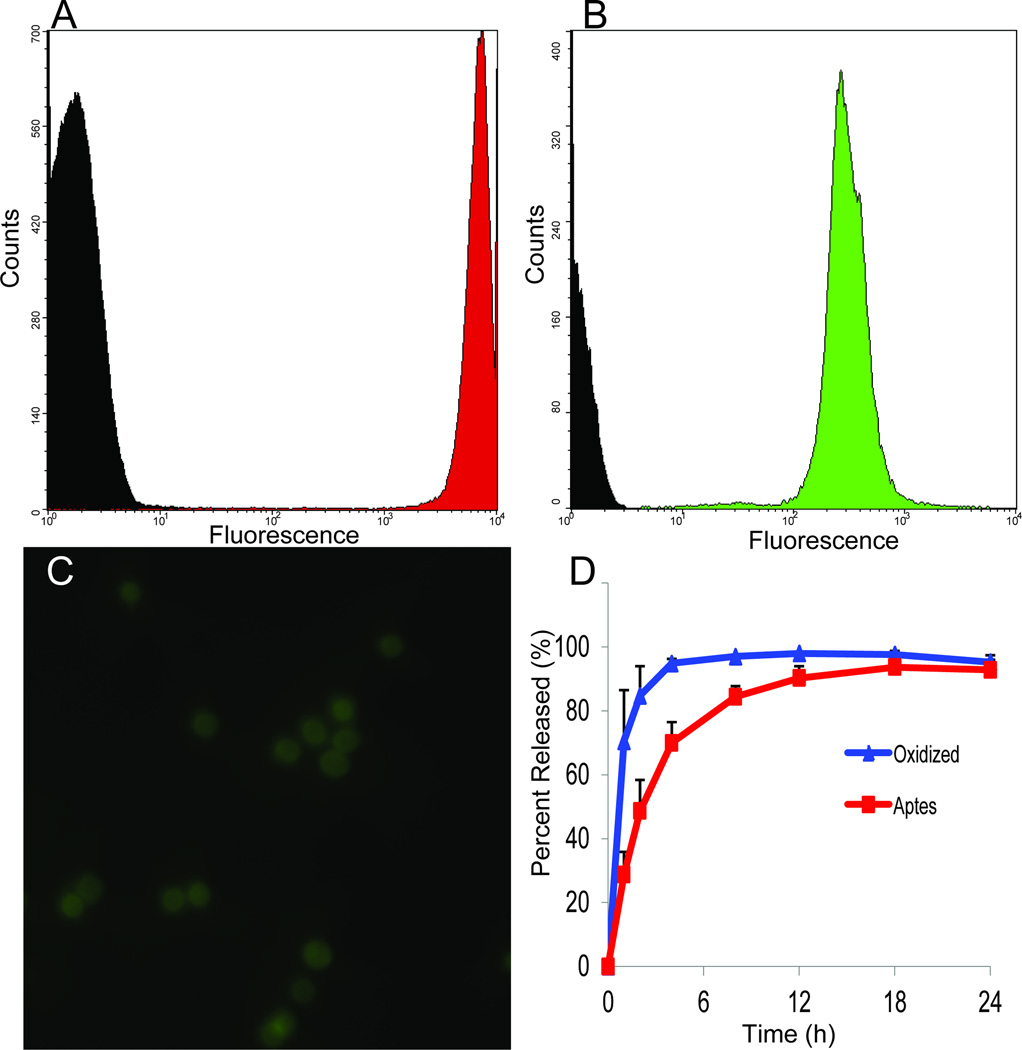
Nanomaterials are advancing in several directions with significant progress being achieved with respect to their synthesis, functionalization and biomedical application. In this review, we will describe several classes of prototypical nanocarriers, such as liposomes, silicon particles, and gold nanoshells, in terms of their individual function as well as their synergistic use. Active and passive targeting, photothermal ablation, and drug controlled release constitute some of the crucial functions identified to achieve a medical purpose. Current limitations in targeting, slow clearance, and systemic as well as local toxicity are addressed in reference to the recent studies that attempted to comprehend and solve these issues. The demand for a more sophisticated understanding of the impact of nanomaterialson the body and of their potential immune response underlies this discussion. Combined components are then discussed in the setting of multifunctional nanocarriers, a class of drug delivery systems we envisioned, proposed, and evolved in the last 5 years. In particular, our third generation of nanocarriers, the multistage vectors, usher in the new field of nanomedicine by combining several components onto multifunctional nanocarriers characterized by emerging properties and able to achieve synergistic effects.
Keywords: cancer; drug delivery; multifunctional; multistage; nanomedicine; nanoparticle; porous silicon; third generation nanocarriers.
Figures
Similar articles
-
Polydopamine-Modified Liposomes: Preparation and Recent Applications in the Biomedical Field.ACS Omega. 2024 May 28;9(23):24105-24120. doi: 10.1021/acsomega.4c02555. eCollection 2024 Jun 11. ACS Omega. 2024. PMID: 38882106 Free PMC article. Review.
-
Silica-Based Nanoparticles for Biomedical Applications: From Nanocarriers to Biomodulators.Acc Chem Res. 2020 Aug 18;53(8):1545-1556. doi: 10.1021/acs.accounts.0c00280. Epub 2020 Jul 15. Acc Chem Res. 2020. PMID: 32667182
-
Nanotechnology: an evidence-based analysis.Ont Health Technol Assess Ser. 2006;6(19):1-43. Epub 2006 Nov 1. Ont Health Technol Assess Ser. 2006. PMID: 23074489 Free PMC article.
-
Multifunctional Nanocarriers for diagnostics, drug delivery and targeted treatment across blood-brain barrier: perspectives on tracking and neuroimaging.Part Fibre Toxicol. 2010 Mar 3;7:3. doi: 10.1186/1743-8977-7-3. Part Fibre Toxicol. 2010. PMID: 20199661 Free PMC article. Review.
-
Targeted multifunctional lipid-based nanocarriers for image-guided drug delivery.Anticancer Agents Med Chem. 2007 Jul;7(4):425-40. doi: 10.2174/187152007781058613. Anticancer Agents Med Chem. 2007. PMID: 17630918 Review.
Cited by
-
The evolution of nucleosidic analogues: self-assembly of prodrugs into nanoparticles for cancer drug delivery.Nanoscale Adv. 2021 Feb 22;3(8):2157-2179. doi: 10.1039/d0na01084g. eCollection 2021 Apr 20. Nanoscale Adv. 2021. PMID: 36133769 Free PMC article. Review.
-
Biomimetic nanoparticles with enhanced affinity towards activated endothelium as versatile tools for theranostic drug delivery.Theranostics. 2018 Jan 5;8(4):1131-1145. doi: 10.7150/thno.22078. eCollection 2018. Theranostics. 2018. PMID: 29464004 Free PMC article.
-
Reversal of EGFR inhibitors' resistance by co-delivering EGFR and integrin αvβ3 inhibitors with nanoparticles in non-small cell lung cancer.Biosci Rep. 2019 Aug 28;39(8):BSR20181259. doi: 10.1042/BSR20181259. Print 2019 Aug 30. Biosci Rep. 2019. PMID: 31316001 Free PMC article.
-
Development and Characterization of PLGA-Based Multistage Delivery System for Enhanced Payload Delivery to Targeted Vascular Endothelium.Macromol Biosci. 2021 Mar;21(3):e2000377. doi: 10.1002/mabi.202000377. Epub 2021 Jan 4. Macromol Biosci. 2021. PMID: 33393217 Free PMC article.
-
Spotlight on Genetic Kidney Diseases: A Call for Drug Delivery and Nanomedicine Solutions.ACS Nano. 2023 Apr 11;17(7):6165-6177. doi: 10.1021/acsnano.2c12140. Epub 2023 Mar 29. ACS Nano. 2023. PMID: 36988207 Free PMC article. Review.
References
-
- Sanvicens N, Marco MP. Multifunctional nanoparticles--properties and prospects for their use in human medicine. Trends Biotechnol. 2008;26:425–433. - PubMed
-
- Babincova M, Altanerova V, Altaner C, et al. In vivo heating of magnetic nanoparticles in alternating magnetic field. Med Phys. 2004;31:2219–2221. - PubMed
-
- Pastan I, Hassan R, Fitzgerald DJ, et al. Immunotoxin therapy of cancer. Nat Rev Cancer. 2006;6:559–565. - PubMed
-
- O'Brien ME, Wigler N, Inbar M, et al. Reduced cardiotoxicity and comparable efficacy in a phase iii trial of pegylated liposomal doxorubicin hcl (caelyx/doxil) versus conventional doxorubicin for first-line treatment of metastatic breast cancer. Ann Oncol. 2004;15:440–449. - PubMed
Grants and funding
LinkOut - more resources
Full Text Sources
Other Literature Sources