

Structural and functional aspects of the Helicobacter pylori secretome
- PMID: 24587618
- PMCID: PMC3925851
- DOI: 10.3748/wjg.v20.i6.1402
Structural and functional aspects of the Helicobacter pylori secretome
Abstract
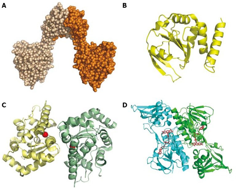
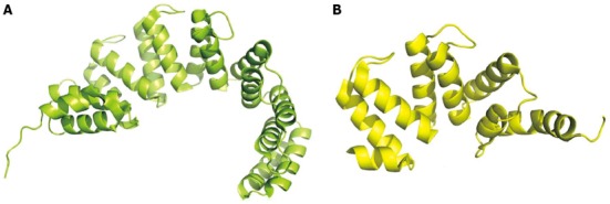
Proteins secreted by Helicobacter pylori (H. pylori), an important human pathogen responsible for severe gastric diseases, are reviewed from the point of view of their biochemical characterization, both functional and structural. Despite the vast amount of experimental data available on the proteins secreted by this bacterium, the precise size of the secretome remains unknown. In this review, we consider as secreted both proteins that contain a secretion signal for the periplasm and proteins that have been detected in the external medium in in vitro experiments. In this way, H. pylori's secretome appears to be composed of slightly more than 160 proteins, but this number must be considered very cautiously, not only because the definition of secretome itself is ambiguous but also because the included proteins were observed as secreted in in vitro experiments that were not representative of the environmental situation in vivo. The proteins that appear to be secreted can be grouped into different classes: enzymes (48 proteins), outer membrane proteins (43), components of flagella (11), members of the cytotoxic-associated genes pathogenicity island or other toxins (8 and 5, respectively), binding and transport proteins (9), and others (11). A final group, which includes 28 members, is represented by hypothetical uncharacterized proteins. Despite the large amount of data accumulated on the H. pylori secretome, a considerable amount of work remains to reach a full comprehension of the system at the molecular level.
Keywords: Helicobacter pylori; Periplasmic space; Secreted proteins; Secretion signal; Secretome.
Figures
References
-
- Marshall BJ, Warren JR. Unidentified curved bacilli in the stomach of patients with gastritis and peptic ulceration. Lancet. 1984;1:1311–1315. - PubMed
-
- Goodwin CS, Mendall MM, Northfield TC. Helicobacter pylori infection. Lancet. 1997;349:265–269. - PubMed
-
- Covacci A, Telford JL, Del Giudice G, Parsonnet J, Rappuoli R. Helicobacter pylori virulence and genetic geography. Science. 1999;284:1328–1333. - PubMed
-
- Tomb JF, White O, Kerlavage AR, Clayton RA, Sutton GG, Fleischmann RD, Ketchum KA, Klenk HP, Gill S, Dougherty BA, et al. The complete genome sequence of the gastric pathogen Helicobacter pylori. Nature. 1997;388:539–547. - PubMed
Publication types
MeSH terms
Substances
LinkOut - more resources
Full Text Sources
Other Literature Sources
Medical
