

Case Reports
doi: 10.3748/wjg.v20.i6.1630.
Characterization of primary hepatic carcinosarcoma by contrast-enhanced ultrasonography: a case report
Affiliations
- PMID: 24587642
- PMCID: PMC3925875
- DOI: 10.3748/wjg.v20.i6.1630
Item in Clipboard
Case Reports
Characterization of primary hepatic carcinosarcoma by contrast-enhanced ultrasonography: a case report
World J Gastroenterol.
.
Abstract
Primary hepatic carcinosarcoma is a rare tumor and is comprised of a mixture of carcinomatous and sarcomatous elements. We present a case of primary carcinosarcoma of the liver in a 59-year-old woman, which was confirmed by pathology following surgical resection. Using contrast-enhanced ultrasonography, the tumor showed peripheral nodular hyperenhancement in the arterial phase with two feeding arterial vessels and a large internal non-enhancing portion in the center. The peripheral nodular portion of the tumor showed hypoenhancement in the later phase.
Keywords: Carcinosarcoma; Contrast-agent; Liver; Ultrasonography.
Figures
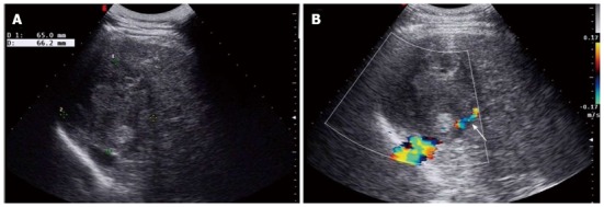
Conventional ultrasonography of a primary hepatic carcinosarcoma. A: Gray-scale ultrasonography showed an ovoid heterogeneous echogenic mass in the right liver; B: Color doppler imaging showed the right hepatic vein compressed by the mass (arrow).
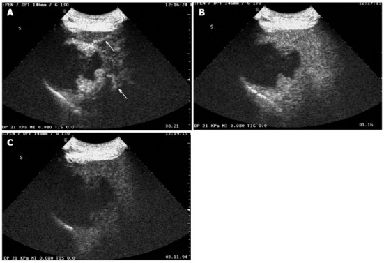
Contrast-enhanced ultrasonography of a primary hepatic carcinosarcoma. A: Contrast-enhanced sonography showed early intense peripheral nodular enhancement in the arterial phase (21 s) with two feeding arteries (arrows); B: Peripheral nodular portion of the tumor was iso-enhancing compared to the surrounding liver in the portal phase (76 s); C: Peripheral nodular portion of the tumor was washed out and was hypo-enhanced compared to the liver in the later phase (191 s).
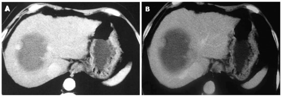
Contrast-enhanced computed tomographic of a primary hepatic carcinosarcoma. A: Contrast-enhanced computed tomographic revealed peripheral nodular enhancement in the arterial phase with a large internal non-enhancing portion, which correlated with contrast-enhanced ultrasonography findings; B: Peripheral nodular portion of the tumor was isoenhanced in the portal phase.
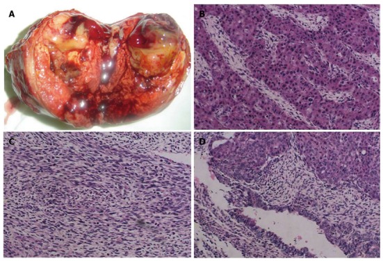
Pathologic findings of the primary hepatic carcinosarcoma (hematoxylin and eosin, original magnification × 200). A: A section of the gross specimen showing a well-demarcated, peripheral nodular solid mass with a central area of necrosis and hemorrhage; B: Light microscopy showing moderate hepatocellular carcinoma; C: Stromal sarcoma components are comprised of spindle cells; D: Atypical spindle and epithelial cells are seen, which are consistent with hepatic carcinosarcoma.
References
-
- Ishak KG, Anthony PP, Sobin LH. Histological typing of tumours of the liver (WHO. World Health Organization. International Histological Classification of Tumours). 2nd ed. Berlin: Springer-Verlag; 1994. pp. 27–28.
-
- Lin YS, Wang TY, Lin JC, Wang HY, Chou KF, Shih SC, Chen MJ. Hepatic carcinosarcoma: clinicopathologic features and a review of the literature. Ann Hepatol. 2013;12:495–500. - PubMed
-
- Aparicio MA, Esteban C, Bengoechea O, Muñoz-Bellvís L. Primary carcinosarcoma of the liver: an unusual case with clearly separated epithelial and mesenchymal components. Rev Esp Enferm Dig. 2011;103:336–338. - PubMed
-
- Celikbilek M, Deniz K, Torun E, Artis T, Ozaslan E, Karahan OI, Patiroglu TE, Ozbakir O. Primary hepatic carcinosarcoma. Hepatobiliary Pancreat Dis Int. 2011;10:101–103. - PubMed
-
- Shu RY, Ye M, Yu WY. A case of primary liver carcinosarcoma: CT findings. Chin J Cancer. 2010;29:346–348. - PubMed
Publication types
MeSH terms
Substances
LinkOut - more resources
Full Text Sources
Other Literature Sources
Medical
