. 2014 Jan 22;48(1):80-6.
doi: 10.2478/raon-2013-0061. eCollection 2014 Mar.

Individual radiosensitivity in a breast cancer collective is changed with the patients' age

Affiliations

Individual radiosensitivity in a breast cancer collective is changed with the patients' age

Judith Auer et al. Radiol Oncol. .

Abstract

Background: Individual radiosensitivity has a crucial impact on radiotherapy related side effects. Our aim was to study a breast cancer collective for its variation of individual radiosensitivity depending on the patients' age.

Materials and methods: Peripheral blood samples were obtained from 129 individuals. Individual radiosensitivity in 67 breast cancer patients and 62 healthy individuals was estimated by 3-color fluorescence in situ hybridization.

Results: Breast cancer patients were distinctly more radiosensitive compared to healthy controls. A subgroup of 9 rather radiosensitive and 9 rather radio-resistant patients was identified. A subgroup of patients aged between 40 and 50 was distinctly more radiosensitive than younger or older patients.

Conclusions: In the breast cancer collective a distinct resistant and sensitive subgroup is identified, which could be subject for treatment adjustment. Preliminary results indicate that especially in the range of age 40 to 50 patients with an increased radiosensitivity are more frequent and may have an increased risk to suffer from therapy related side effects.

Keywords: age; breast cancer; chromosomal aberrations; fluorescence in situ hybridization; individual radiosensitivity; radiotherapy.


Figures

FIGURE 1:.
FIGURE 1:.
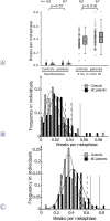
A – Three-color FISH painting of chromosomes 1 (red), 2 (green) and 4 (yellow). A metaphase without aberrations (left) and a metaphase (right) with a reciprocal dicentric chromosome and a break are displayed. The two aberrations were scored as 3 breaks. B – Frequency of patients and controls age distribution.
FIGURE 2.
FIGURE 2.
Individual chromosomal radiosensitivity as measured in in vitro and in vivo irradiated lymphocytes by three-color FISH. Chromosomal aberrations were scored as breaks per metaphase. Blood samples were derived from healthy individuals (controls) and breast cancer patients (BC patients). A – Breaks per metaphase of unirradiated samples (spontaneous) and after in vitro irradiation by a dose of 2 Gy (2 Gy in vitro IR) were shown as scatter plots and box plots. Frequency of breaks per metaphase fitted to a Gaussian distributions of B – unirradiated samples, C - after in vitro irradiation by a dose of 2 Gy
FIGURE 3.
FIGURE 3.
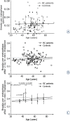
Breaks per metaphase in dependence of the patients (open square, continuous line) and healthy individuals age (filled triangle, dashed line). Ten year intervals were summarized and the average was given as mean value with its standard deviation. A – Unirradiated blood samples, B – samples after 2 Gy in vitro irradiation individual values and C – samples after 2 Gy in vitro irradiation mean values and standard deviation. B C – Thin line gives the linear regression including all patients values, thick continuous line gives the linear regression excluding the group aged 40 to 50 years.
FIGURE 4.
FIGURE 4.
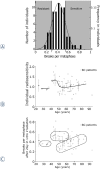
A - Classification of the breast cancer patients on the basis of the breaks per metaphase level after in vivo irradiation (2 Gy IR). B – Putative individual radiosensitivity dependent on breast cancers patients’ age. C - Presumed classification of the breast cancer patients in three groups of different radiosensitivity.

References

    1. Huber R, Braselmann H, Geinitz H, Jaehnert J, Baumgartner A, Thamm R, et al. Chromosomal radiosensitivity and acute radiation side effects after radiotherapy in tumour patients--a follow-up study. Radiat Oncol. 2011;6:32. - PMC - PubMed
    1. Dikomey E, Borgmann K, Peacock J, Jung H. Why recent studies relating normal tissue response to individual radiosensitivity might have failed and how new studies should be performed. Int J Radiat Oncol Biol Phys. 2003;56:1194–200. - PubMed
    1. Zschenker O, Raabe A, Boeckelmann IK, Borstelmann S, Szymczak S, Wellek S, et al. Association of single nucleotide polymorphisms in ATM, GSTP1, SOD2, TGFB1, XPD and XRCC1 with clinical and cellular radiosensitivity. Radiother Oncol. 2010;97:26–32. - PubMed
    1. Lopez E, Guerrero R, Nunez MI, del Moral R, Villalobos M, Martinez-Galan J, et al. Early and late skin reactions to radiotherapy for breast cancer and their correlation with radiation-induced DNA damage in lymphocytes. Breast Cancer Res. 2005;7:R690–8. - PMC - PubMed
    1. Ambrosone CB, Tian C, Ahn J, Kropp S, Helmbold I, von Fournier D, et al. Genetic predictors of acute toxicities related to radiation therapy following lumpectomy for breast cancer: a case-series study. Breast Cancer Res. 2006;8:R40. - PMC - PubMed

LinkOut - more resources