Global small-angle X-ray scattering data analysis for multilamellar vesicles: the evolution of the scattering density profile model
- PMID: 24587787
- PMCID: PMC3937811
- DOI: 10.1107/S1600576713029798
Global small-angle X-ray scattering data analysis for multilamellar vesicles: the evolution of the scattering density profile model
Abstract
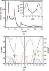
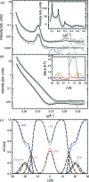
The highly successful scattering density profile (SDP) model, used to jointly analyze small-angle X-ray and neutron scattering data from unilamellar vesicles, has been adapted for use with data from fully hydrated, liquid crystalline multilamellar vesicles (MLVs). Using a genetic algorithm, this new method is capable of providing high-resolution structural information, as well as determining bilayer elastic bending fluctuations from standalone X-ray data. Structural parameters such as bilayer thickness and area per lipid were determined for a series of saturated and unsaturated lipids, as well as binary mixtures with cholesterol. The results are in good agreement with previously reported SDP data, which used both neutron and X-ray data. The inclusion of deuterated and non-deuterated MLV neutron data in the analysis improved the lipid backbone information but did not improve, within experimental error, the structural data regarding bilayer thickness and area per lipid.
Keywords: genetic algorithms; liquid crystalline multilamellar vesicles; scattering density profile model; small-angle X-ray scattering.
Figures


Similar articles
-
Structural and mechanical properties of cardiolipin lipid bilayers determined using neutron spin echo, small angle neutron and X-ray scattering, and molecular dynamics simulations.Soft Matter. 2015 Jan 7;11(1):130-8. doi: 10.1039/c4sm02227k. Soft Matter. 2015. PMID: 25369786
-
Erratum: Neutron Spin Echo Spectroscopy as a Unique Probe for Lipid Membrane Dynamics and Membrane-Protein Interactions.J Vis Exp. 2021 Aug 6;(174). doi: 10.3791/6475. J Vis Exp. 2021. PMID: 34358222
-
Joint small-angle X-ray and neutron scattering data analysis of asymmetric lipid vesicles.J Appl Crystallogr. 2017 Feb 28;50(Pt 2):419-429. doi: 10.1107/S1600576717000656. eCollection 2017 Apr 1. J Appl Crystallogr. 2017. PMID: 28381971 Free PMC article.
-
Model-based approaches for the determination of lipid bilayer structure from small-angle neutron and X-ray scattering data.Eur Biophys J. 2012 Oct;41(10):875-90. doi: 10.1007/s00249-012-0817-5. Epub 2012 May 16. Eur Biophys J. 2012. PMID: 22588484 Review.
-
Small-Angle Neutron Scattering for Studying Lipid Bilayer Membranes.Biomolecules. 2022 Oct 29;12(11):1591. doi: 10.3390/biom12111591. Biomolecules. 2022. PMID: 36358941 Free PMC article. Review.
Cited by
-
All n-3 PUFA are not the same: MD simulations reveal differences in membrane organization for EPA, DHA and DPA.Biochim Biophys Acta Biomembr. 2018 May;1860(5):1125-1134. doi: 10.1016/j.bbamem.2018.01.002. Epub 2018 Jan 3. Biochim Biophys Acta Biomembr. 2018. PMID: 29305832 Free PMC article.
-
Membrane remodeling and mechanics: Experiments and simulations of α-Synuclein.Biochim Biophys Acta. 2016 Jul;1858(7 Pt B):1594-609. doi: 10.1016/j.bbamem.2016.03.012. Epub 2016 Mar 10. Biochim Biophys Acta. 2016. PMID: 26972046 Free PMC article. Review.
-
Restoring structural parameters of lipid mixtures from small-angle X-ray scattering data.J Appl Crystallogr. 2021 Feb 1;54(Pt 1):169-179. doi: 10.1107/S1600576720015368. eCollection 2021 Feb 1. J Appl Crystallogr. 2021. PMID: 33833646 Free PMC article.
-
In vivo analysis of the Escherichia coli ultrastructure by small-angle scattering.IUCrJ. 2017 Sep 26;4(Pt 6):751-757. doi: 10.1107/S2052252517013008. eCollection 2017 Nov 1. IUCrJ. 2017. PMID: 29123677 Free PMC article.
-
Phenylalanine and Tryptophan-Based Surfactants as New Antibacterial Agents: Characterization, Self-Aggregation Properties, and DPPC/Surfactants Vesicles Formulation.Pharmaceutics. 2023 Jun 30;15(7):1856. doi: 10.3390/pharmaceutics15071856. Pharmaceutics. 2023. PMID: 37514042 Free PMC article.
References
-
- Caillé, A. (1972). C. R. Acad. Sci. Paris B, 274, 891–893.
-
- Frühwirth, T., Fritz, G., Freiberger, N. & Glatter, O. (2004). J. Appl. Cryst. 37, 703–710.
-
- Goldberg, D. (1989). Genetic Algorithms in Search, Optimization, and Machine Learning. New York: Addison-Wesley Professional.
Grants and funding
LinkOut - more resources
Full Text Sources
Other Literature Sources
