Review
. 2014:2014:581468.
doi: 10.1155/2014/581468. Epub 2014 Jan 23.

A New Diagnostic Way for Behcet's Disease: Skin Prick with Self-Saliva

Affiliations
Review

A New Diagnostic Way for Behcet's Disease: Skin Prick with Self-Saliva

Fumio Kaneko et al. Genet Res Int. 2014.

Abstract

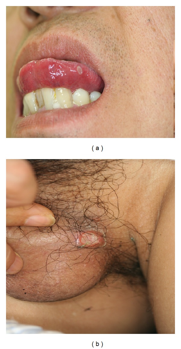
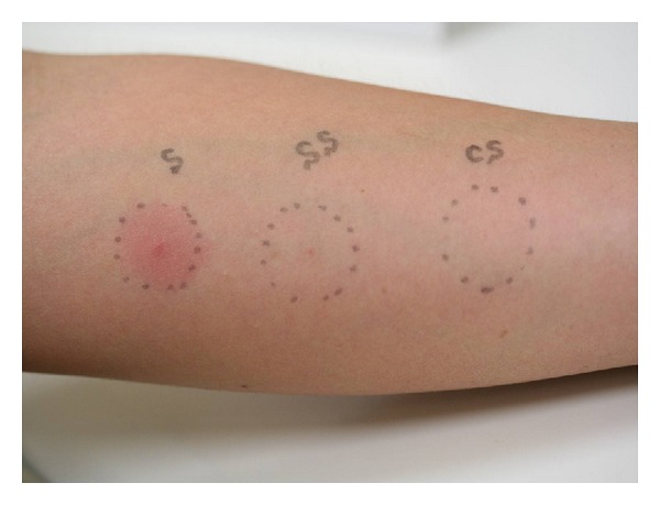
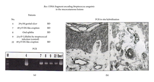
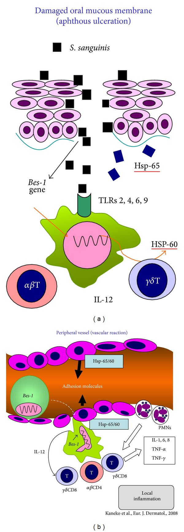
Behcet's disease (BD) is a mysterious multisystemic disorder characterized by recurrent involvement of mucocutaneous (including recurrent aphthous stomatitis; RAS), ocular, intestinal, vascular, and/or nervous system organs. Previously, the positivity of "pathergy test", which is one of the diagnostic examinations, was reported to be related to the possession of HLA-B51 gene in BD patients, even though the positivity is low and different from the countries. Here, instead of the ordinal pathergy test, we would like to propose the prick with self-saliva as a new diagnostic way for patients with RAS of BD based on the genetic intrinsic factors including HLA-B51 and extrinsic triggering factors. BD patients are considered to acquire the hypersensitivity against oral streptococci through the innate immune mechanism in the oral cavity. Bes-1 gene and 65 kD of heat shock protein (HSP-65) derived from oral S. sanguinis are supposed to play important roles as extrinsic factors in BD pathogenesis. Although the prick positivity was not related to the possession of HLA-B51 gene, the method is suggested to be a significant way for BD diagnosis. The results also suggest that BD symptoms are due to the vascular immune responses by monocytes expressed oral streptococcal agents of the patients.


Figures

Figure 1
Figure 1
Oral aphtha and genital ulcerations seen in a male BD patient with neuropathy (55 M YY in Table 1). (a) Oral ulcer round and punched-out shaped on the tongue. (b) Genital ulcer shaped like oral ulcer.
Figure 2
Figure 2
Histology of aphthous ulceration of a BD patient. (a) Aphthous ulcer of the lip defecting the epithelial layer (HE, ×100). (b) Magnified feature of the ulcer edge of the epithelial layer. The epithelial cells are surrounded by inflammatory infiltrates like “Rosetta formation.”
Figure 3
Figure 3
EN-like eruption and the histology and immunohistology. (a) EN-like eruption on the lower legs of a BD patient. (b) Vasculitis infiltrated by lymphoid cells and neutrophils (HE, 400x). (c) Deposits of streptococcal antigen adhering to the vascular wall (direct immunofluorescence, ×400).
Figure 4
Figure 4
Incubation of saliva of a BD patient using MS (mitis and salivarius) agar in which oral streptococci are selectively grown. (a) Oral streptococci grew from saliva in 5 day. (b) Area of sterilized saliva using syringe micromembrane filter.
Figure 5
Figure 5
Prick test with self-saliva using Lancetter (33 F AT in Table 1). The skin reactions were observed 48 hours after prick. S: self-saliva; SS: sterilized saliva using syringe-filter with 0.2 μm pores; CS: control saline.
Figure 6
Figure 6
Bes-1 gene expression in the mucocutaneous lesions of patients with Behcet's disease (BD) [43]. (a) Three of 11 BD patients were positive for Bes-1 DNA in the lesions including aphthous and genital ulcerations and erythema nodosum (EN)-like eruption by amplified polymerase chain reaction (PCR) using the primers: Bes-1-1 (5′-TAATAACCCTGACCAAGCCTA-3′) and Bes-1-2 (5′-CCCTTTCAAAAGTCATAAATC-3′) encoding S. sanguinis. (b) In these positive lesions, Bes-1 DNA was also detected in the cytoplasm of monocytes adhering to the vascular walls and infiltrated around the vessels by PCR in situ hybridization.
Figure 7
Figure 7
Hypothesis of the mechanisms in the appearance of various lesions of BD patients [49, 50]. (a) The antigen presenting cells (APCs) (macrophages and/or dendritic cells) immunized by S. sanguinis agents though TLRs in the oral cavity might be carried to the peripheral regions. (b) If the APCs in the blood flow adhered to the impaired and/or MICA and adhesion molecules expressed endothelial cells of vascular wall, the immunological reaction might be appeared as BD lesion.

References

    1. Behcet H. Uber rezidivierende, aphthous durch ein Virus verursachte Geschwuere am Mund, am Auge und an den Genitalien. Dermatologische Wochenschrift. 1937;105:1152–1157.
    1. Altenburg A, Papoutsis N, Orawa H, Martus P, Krause L, Zouboulis CC. Epidemiology and clinical manifestations of Adamantiades-Behcet disease in Germany—current pathogenetic concepts and therapeutic possibilities. Journal of the German Society of Dermatology. 2006;4(1):49–66. - PubMed
    1. Alpsoy E, Zouboulis C, Ehrlich GE. Mucocutaneous lesions of Behcet’s disease. Yonsei Medical Journal. 2007;48(4):573–585. - PMC - PubMed
    1. Ohno S, Onguchi M, Hirose S, Matsuda H, Wakisaka A, Aizawa M. Close association of HLA-Bw51 with Behcet’s disease. Archives of Ophthalmology. 1982;100(9):1455–1458. - PubMed
    1. Zouboulis CC, May T. Pathogenesis of Adamantiades-Behcet’s disease. Medical Microbiology and Immunology. 2003;192(3):149–155. - PubMed

LinkOut - more resources