

p16 Expression Is Lost in Severely Atypical Cellular Blue Nevi and Melanoma Compared to Conventional, Mildly, and Moderately Atypical Cellular Blue Nevi
- PMID: 24587914
- PMCID: PMC3920610
- DOI: 10.1155/2014/348417
p16 Expression Is Lost in Severely Atypical Cellular Blue Nevi and Melanoma Compared to Conventional, Mildly, and Moderately Atypical Cellular Blue Nevi
Abstract
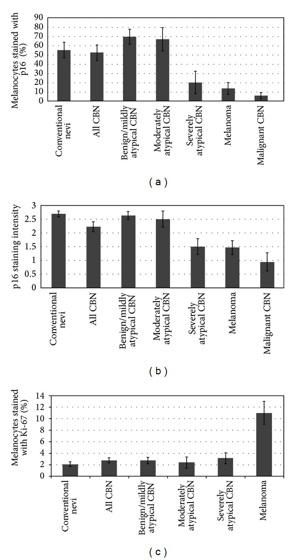
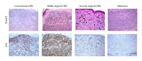
Background. Significant decreases in p16 expression have been shown to occur in melanoma compared to Spitz tumors, and loss of p16 staining has been found to correlate with melanoma tumor progression. However, comparison of p16 between atypical cellular blue nevi (CBN) and melanoma has not been reported previously. Methods. p16 immunohistochemical staining was evaluated in 14 atypical CBN, 8 conventional and atypical melanocytic nevi, and 16 melanomas, including 4 malignant CBN. p16 staining intensity was graded on a scale of 0-3 and the percentage of melanocytes stained with p16 was determined. Results. p16 staining was significantly higher in all CBN as a group when compared to melanomas (P = 0.001) and malignant CBN (P = 0.00008). Higher p16 expression was also seen in mildly (P = 0.0002) and moderately atypical (P = 0.02), but not severely atypical, CBN compared to melanomas. Conclusions. p16 immunohistochemical expression is higher in mildly and moderately atypical CBN compared to severely atypical CBN and melanomas. In conjunction with additional markers and histology, p16 staining may be useful in confirming the benign nature of these tumors, but is not useful in distinguishing severely atypical CBN from malignant cases, consistent with the overlapping histologic features between these tumors.
Figures
Similar articles
-
Atypical cellular blue nevi (cellular blue nevi with atypical features): lack of consensus for diagnosis and distinction from cellular blue nevi and malignant melanoma ("malignant blue nevus").Am J Surg Pathol. 2008 Jan;32(1):36-44. doi: 10.1097/PAS.0b013e3181573aaf. Am J Surg Pathol. 2008. PMID: 18162768
-
Cyclin D1 and p16 Expression in Blue Nevi and Malignant Melanoma.Appl Immunohistochem Mol Morphol. 2017 Feb;25(2):91-94. doi: 10.1097/PAI.0000000000000276. Appl Immunohistochem Mol Morphol. 2017. PMID: 26766120
-
p16 expression: a marker of differentiation between childhood malignant melanomas and Spitz nevi.J Am Acad Dermatol. 2011 Aug;65(2):357-363. doi: 10.1016/j.jaad.2010.07.031. Epub 2011 May 6. J Am Acad Dermatol. 2011. PMID: 21550132
-
Atypical Spitz Tumors: A Diagnostic Challenge.Arch Pathol Lab Med. 2015 Oct;139(10):1263-70. doi: 10.5858/arpa.2015-0207-RA. Arch Pathol Lab Med. 2015. PMID: 26414472 Review.
-
An update on molecular alterations in melanocytic tumors with emphasis on Spitzoid lesions.Ann Transl Med. 2018 Jun;6(12):249. doi: 10.21037/atm.2018.05.23. Ann Transl Med. 2018. PMID: 30069451 Free PMC article. Review.
Cited by
-
A p16-Ki-67-HMB45 immunohistochemistry scoring system as an ancillary diagnostic tool in the diagnosis of melanoma.Diagn Pathol. 2015 Oct 26;10:195. doi: 10.1186/s13000-015-0431-9. Diagn Pathol. 2015. PMID: 26503349 Free PMC article.
-
Epigenetic effects of cadmium in cancer: focus on melanoma.Curr Genomics. 2014 Dec;15(6):420-35. doi: 10.2174/138920291506150106145932. Curr Genomics. 2014. PMID: 25646071 Free PMC article.
-
Immune-phenotypical markers for the differential diagnosis of melanocytic lesions.Int J Clin Exp Pathol. 2015 Sep 1;8(9):9742-51. eCollection 2015. Int J Clin Exp Pathol. 2015. PMID: 26617684 Free PMC article. Review.
-
Cellular Blue Nevus Diagnosed following Excision of Melanoma: A Challenge in Diagnosis.Case Rep Pathol. 2016;2016:8107671. doi: 10.1155/2016/8107671. Epub 2016 May 26. Case Rep Pathol. 2016. PMID: 27313934 Free PMC article.
References
-
- Ruiter DJ, van Dijk MCRF, Ferrier CM. Current diagnostic problems in melanoma pathology. Seminars in Cutaneous Medicine and Surgery. 2003;22(1):33–41. - PubMed
-
- Brochez L, Verhaeghe E, Grosshans E, et al. Inter-observer variation in the histopathological diagnosis of clinically suspicious pigmented skin lesions. Journal of Pathology. 2002;196(4):459–466. - PubMed
-
- Tom WL, Hsu JW, Eichenfield LF, Friedlander SF. Pediatric “sTUMP” lesions: evaluation and management of difficult atypical Spitzoid lesions in children. Journal of the American Academy of Dermatology. 2011;64(3):559–572. - PubMed
-
- Barnhill RL, Argenyi Z, Berwick M, et al. Atypical cellular blue nevi (cellular blue nevi with atypical features): lack of consensus for diagnosis and distinction from cellular blue nevi and malignant melanoma (“malignant blue nevus”) The American Journal of Surgical Pathology. 2008;32(1):36–44. - PubMed
-
- Murali R, McCarthy SW, Scolyer RA. Blue nevi and related lesions: a review highlighting atypical and newly described variants, distinguishing features and diagnostic pitfalls. Advances in Anatomic Pathology. 2009;16(6):365–382. - PubMed
LinkOut - more resources
Full Text Sources
Other Literature Sources
