Receptor Interacting Protein 2 (RIP2) Is Dispensable for OVA-Induced Airway Inflammation in Mice
- PMID: 24587954
- PMCID: PMC3936046
- DOI: 10.4168/aair.2014.6.2.163
Receptor Interacting Protein 2 (RIP2) Is Dispensable for OVA-Induced Airway Inflammation in Mice
Abstract
Purpose: Asthma is a pulmonary chronic inflammatory disease characterized by airway obstruction and hyperresponsiveness. Pattern recognition receptors are known to play a key role in the development of allergic diseases as well as host defenses against microbial infection. Receptor interacting protein 2 (RIP2), a serine/threonine kinase, is an adaptor molecule of NOD1 and NOD2, and genetic variation in this receptor is known to be associated with the severity of allergic asthma in children. In this study, we examined the role of RIP2 in the development of allergic airway inflammation in a mouse model.
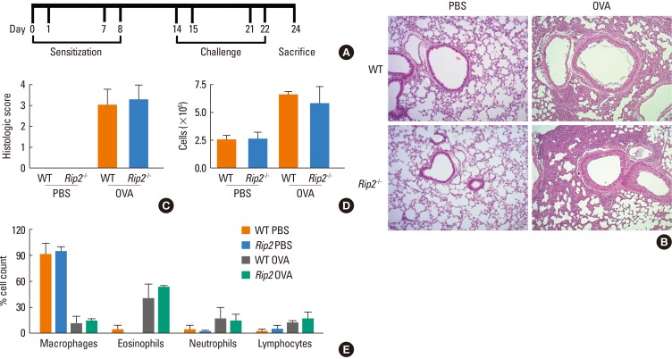
Methods: Airway inflammation was induced in mice through intranasal administration of ovalbumin (OVA) after 2 intraperitoneal immunizations with OVA. Lung inflammation and mucus hypersecretion were examined histologically and total cell infiltration in bronchoalveolar (BAL) fluids was determined. Levels of the Th2-related cytokines, IL-5 and IL-13, in lung extracts were measured by ELISA. Serum antigen-specific IgE and IgG1 levels were also assessed.
Results: OVA-induced lung inflammation and mucus hypersecretion were not different between WT and RIP2-deficient mice. The IL-5 and IL-13 levels in the bronchoalveolar (BAL) fluids were also not impaired in RIP2-deficient mice compared to WT mice. Moreover, RIP2 deficiency did not affect serum OVA-specific IgG1 and IgE levels.
Conclusions: Our results suggest that RIP2 is not associated with the development of allergic airway inflammation.
Keywords: IgE; RIP2; Th2; airway inflammation; ovalbumin.
Conflict of interest statement
There are no financial or other issues that might lead to conflict of interest.
Figures


Similar articles
-
Receptor-interacting protein 2 gene silencing attenuates allergic airway inflammation.J Immunol. 2013 Sep 1;191(5):2691-9. doi: 10.4049/jimmunol.1202416. Epub 2013 Aug 5. J Immunol. 2013. PMID: 23918989
-
The involvement of type 1a angiotensin II receptors in the regulation of airway inflammation in a murine model of allergic asthma.Clin Exp Allergy. 2007 Nov;37(11):1720-7. doi: 10.1111/j.1365-2222.2007.02815.x. Epub 2007 Sep 17. Clin Exp Allergy. 2007. PMID: 17877756
-
TRIF Deficiency does not Affect Severity of Ovalbumin-induced Airway Inflammation in Mice.Immune Netw. 2014 Oct;14(5):249-54. doi: 10.4110/in.2014.14.5.249. Epub 2014 Oct 22. Immune Netw. 2014. PMID: 25360076 Free PMC article.
-
Experimental protocol for development of adjuvant-free murine chronic model of allergic asthma.J Immunol Methods. 2019 May;468:10-19. doi: 10.1016/j.jim.2019.03.002. Epub 2019 Mar 14. J Immunol Methods. 2019. PMID: 30880263
-
Bupleurum chinense extract ameliorates an OVA-induced murine allergic asthma through the reduction of the Th2 and Th17 cytokines production by inactivation of NFκB pathway.Biomed Pharmacother. 2017 Jul;91:1085-1095. doi: 10.1016/j.biopha.2017.04.133. Epub 2017 May 16. Biomed Pharmacother. 2017. PMID: 28531919
Cited by
-
Allergic asthma: RIPK2 takes the lead.J Leukoc Biol. 2018 Sep;104(3):441-443. doi: 10.1002/JLB.3CE0718-293. Epub 2018 Aug 14. J Leukoc Biol. 2018. PMID: 30106490 Free PMC article. No abstract available.
-
Frontline Science: RIP2 promotes house dust mite-induced allergic airway inflammation.J Leukoc Biol. 2018 Sep;104(3):447-459. doi: 10.1002/JLB.4HI0118-017RR. Epub 2018 Jul 27. J Leukoc Biol. 2018. PMID: 30052281 Free PMC article.
-
NOD2 protects against allergic lung inflammation in obese female mice.iScience. 2024 Oct 11;27(11):111130. doi: 10.1016/j.isci.2024.111130. eCollection 2024 Nov 15. iScience. 2024. PMID: 39507249 Free PMC article.
-
Chinese herbal component, Praeruptorin E, enhances anti-asthma efficacy and prevents toxicity of aminophylline by targeting the NF-κB/PXR/CYP3A4 pathway.Ann Transl Med. 2022 Feb;10(4):225. doi: 10.21037/atm-22-386. Ann Transl Med. 2022. PMID: 35280431 Free PMC article.
-
Administration of Pigment Epithelium-Derived Factor Inhibits Airway Inflammation and Remodeling in Chronic OVA-Induced Mice via VEGF Suppression.Allergy Asthma Immunol Res. 2016 Mar;8(2):161-9. doi: 10.4168/aair.2016.8.2.161. Epub 2015 Nov 2. Allergy Asthma Immunol Res. 2016. PMID: 26739410 Free PMC article.
References
-
- Masoli M, Fabian D, Holt S, Beasley R Global Initiative for Asthma (GINA) Program. The global burden of asthma: executive summary of the GINA Dissemination Committee report. Allergy. 2004;59:469–478. - PubMed
-
- Busse WW, Lemanske RF., Jr Asthma. N Engl J Med. 2001;344:350–362. - PubMed
-
- Wenzel SE. Asthma phenotypes: the evolution from clinical to molecular approaches. Nat Med. 2012;18:716–725. - PubMed
LinkOut - more resources
Full Text Sources
Other Literature Sources