

Review
doi: 10.1021/cr400546e.
Epub 2014 Mar 3.
Fluorescent sensors for measuring metal ions in living systems
Affiliations
- PMID: 24588137
- PMCID: PMC4096685
- DOI: 10.1021/cr400546e
Item in Clipboard
Review
Fluorescent sensors for measuring metal ions in living systems
Chem Rev.
.
No abstract available
Figures
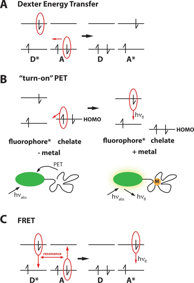
Schematic of Dexter energy transfer (A), “turn-on”
PET (B), and FRET (C).
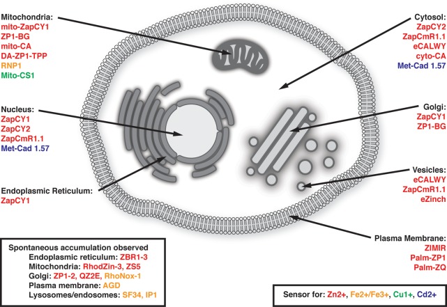
Diagram of sensors that have been targeted to specific organelles
for subcellular metal ion imaging, either with peptide signaling motifs
or with chemical groups known to associate with a particular subcellular
location. Additionally, probes for which spontaneous accumulation
in an organelle has been verified by colocalization studies are shown.
More detailed descriptions of particular targeting strategies are
discussed in later sections.

Timeline of historical
developments in visualizing metal ions in
cells.
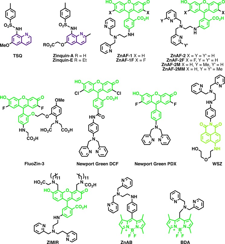
Quinoline-,
fluorescein-, 4-aminonapthalimide-, and BODIPY-based
Zn2+ sensors.
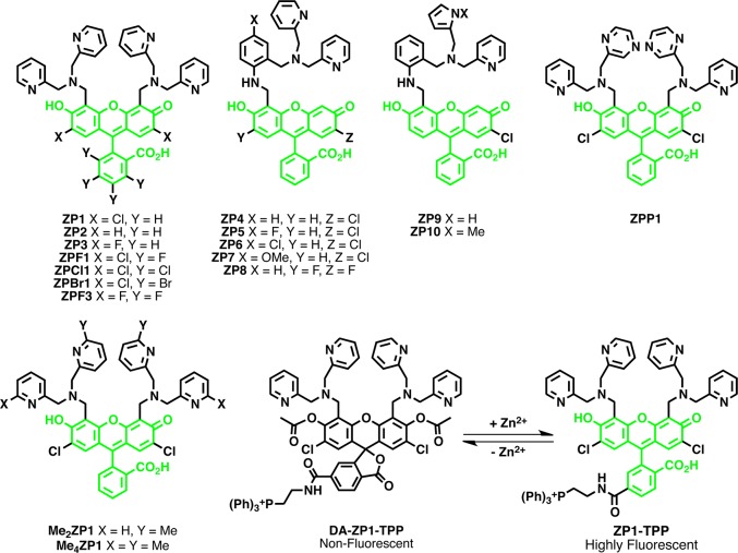
The ZP family of Zn2+ sensors.
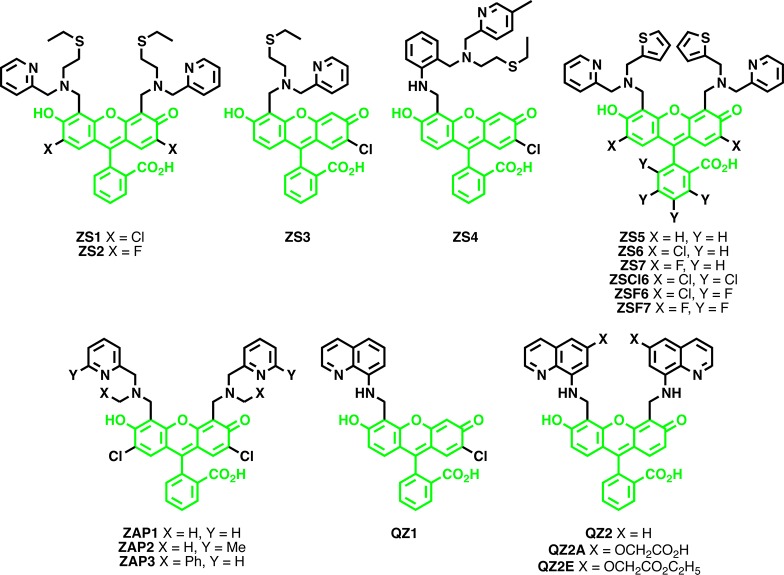
The ZS, QZ, and ZAP families of Zn2+ sensors.
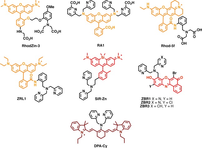
Rhodamine-, resorufin-,
and cyanine-based small-molecule Zn2+ sensors.
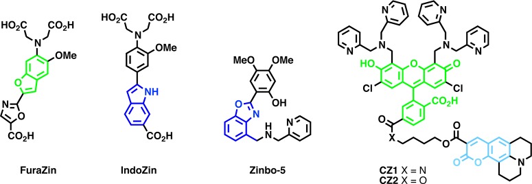
Ratiometric small-molecule Zn2+ sensors.
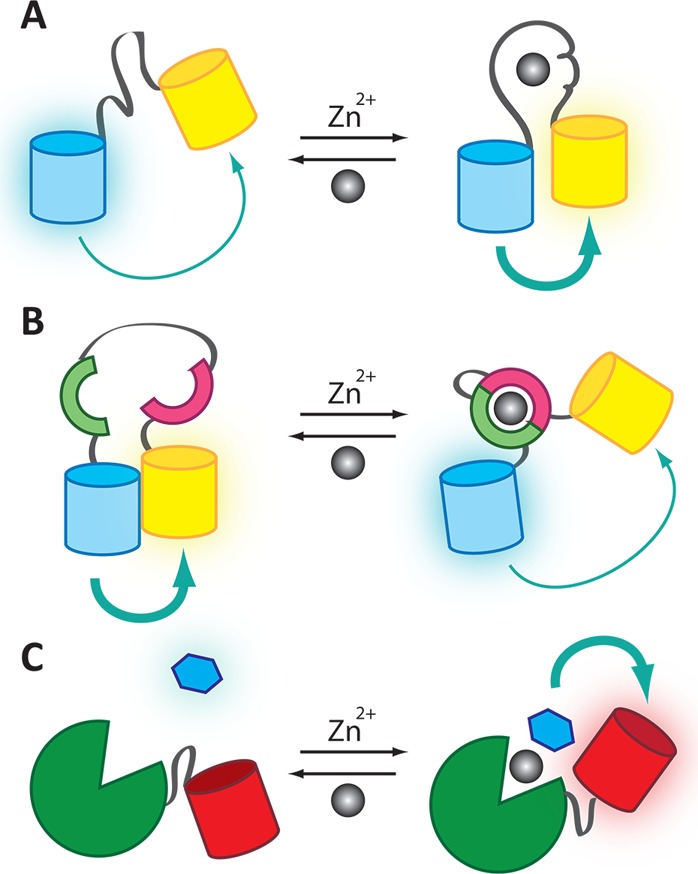
Mechanisms
of metal ion sensing by genetically encoded and hybrid
probes for Zn2+. (A) The Zap and Zif families consist of
one or two Zn2+-finger domains between two FPs. Zn2+ binding induces a conformational change in the Zn2+-finger that leads to a change in FRET ratio. (B) The eCALWY family
uses Zn2+ binding domains from Atox1 and WD4. The two FPs
associate in the absence of Zn2+, but Zn2+ binding
causes association of the binding domains and reduces the FRET efficiency.
(C) The hybrid probe CA-FP has an FP linked to CA. When Zn2+ binds to CA, an exogenously added dapoxyl sulfonamide (blue hexagon)
can bind to an open site on the Zn2+ ion, leading to a
FRET response between the small-molecule fluorophore and the FP.
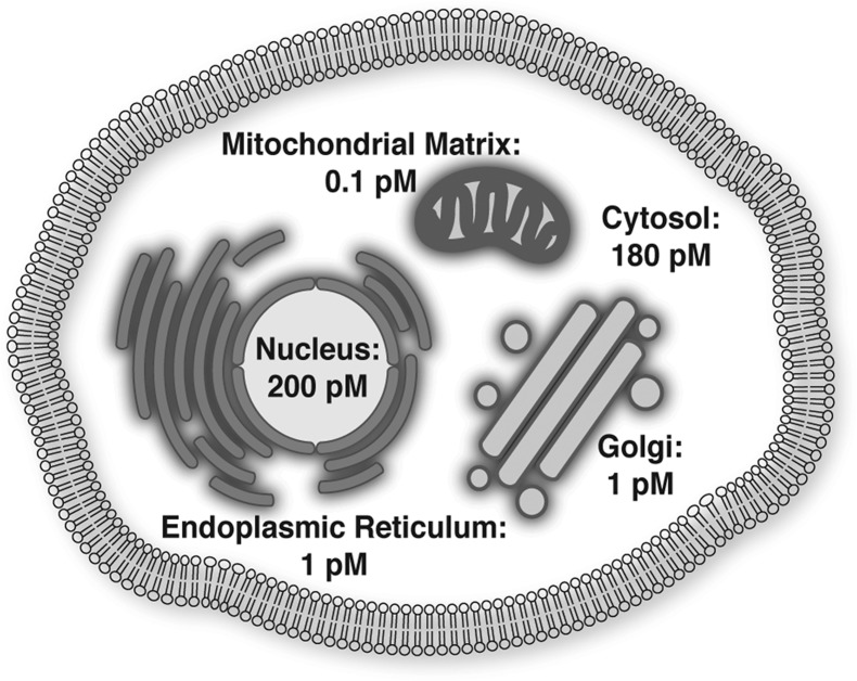
Heterogeneous distribution of Zn2+ throughout the mammalian
cell. Genetically encoded sensors can be targeted to specific compartments
with a signaling sequence to selectively monitor the Zn2+ pool of that organelle.
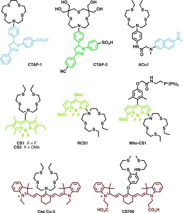
Molecular Cu+ sensors.
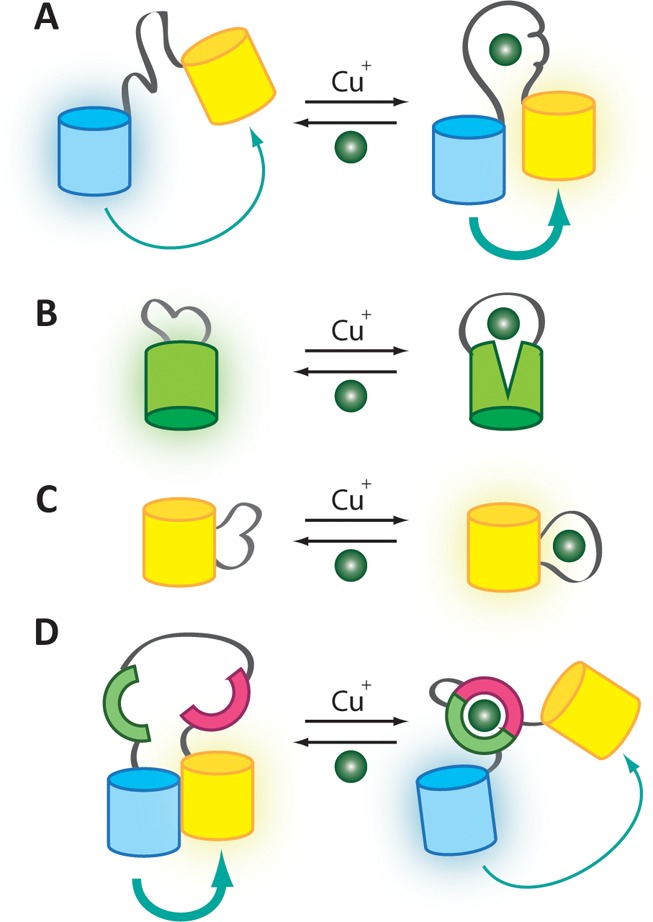
Mechanism of metal ion sensing by genetically
encoded probes for
Cu+. (A) AMT1-FRET, Ace1-FRET, and Mac1-FRET have a cysteine-rich
Cu+-binding domain between a CFP/YFP FRET pair such that
metal binding results in an increased FRET signal. (B) Cu+ binding to EGFP-Amt1 distorts the β-barrel of EGFP and decreases
fluorescence. (C) YFP-Ace1 and related sensors have the Cu+-binding domain of Ace1 inserted between two strands of EYFP. Cu+ binding alters the local environment of the chromophore and
leads to an increased fluorescent signal. (D) The eCALWY Zn2+ sensor platform can be tuned for improved selectivity toward Cu+. In the absence of Cu+, association between two
FPs produces a FRET signal. CU+-induced association between
the metal binding domains of Atox1 and WD4 changes the structure of
the sensor and results in a decreased FRET signal.
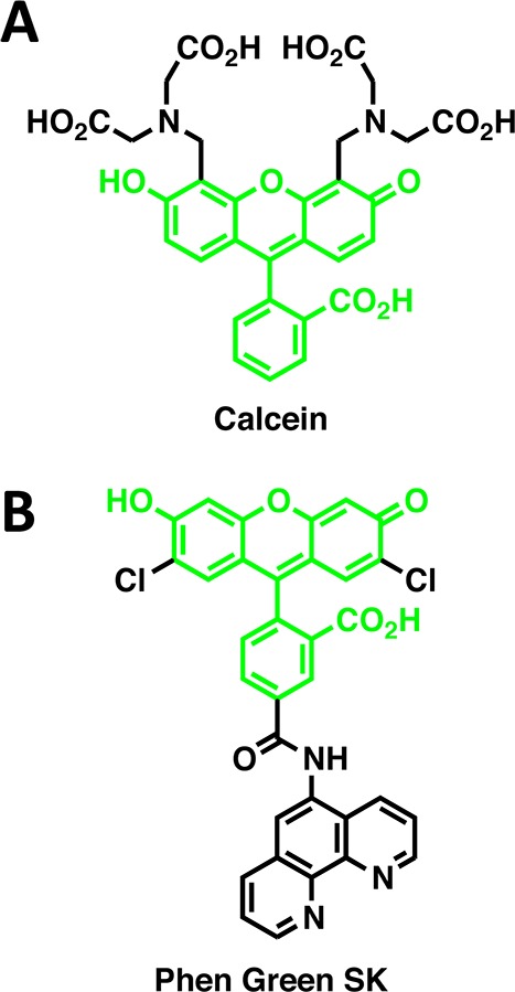
Calcein (A) and Phen Green SK (B) represent
early fluorescent tools
for visualizing cellular iron homeostasis.

Small-molecule Fe2+ sensors.
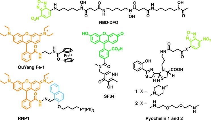
Small-molecule Fe3+ sensors.
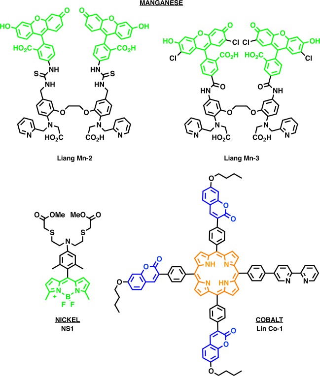
Molecular sensors for the biological metals Mn2+, Ni2+, and Co2+.
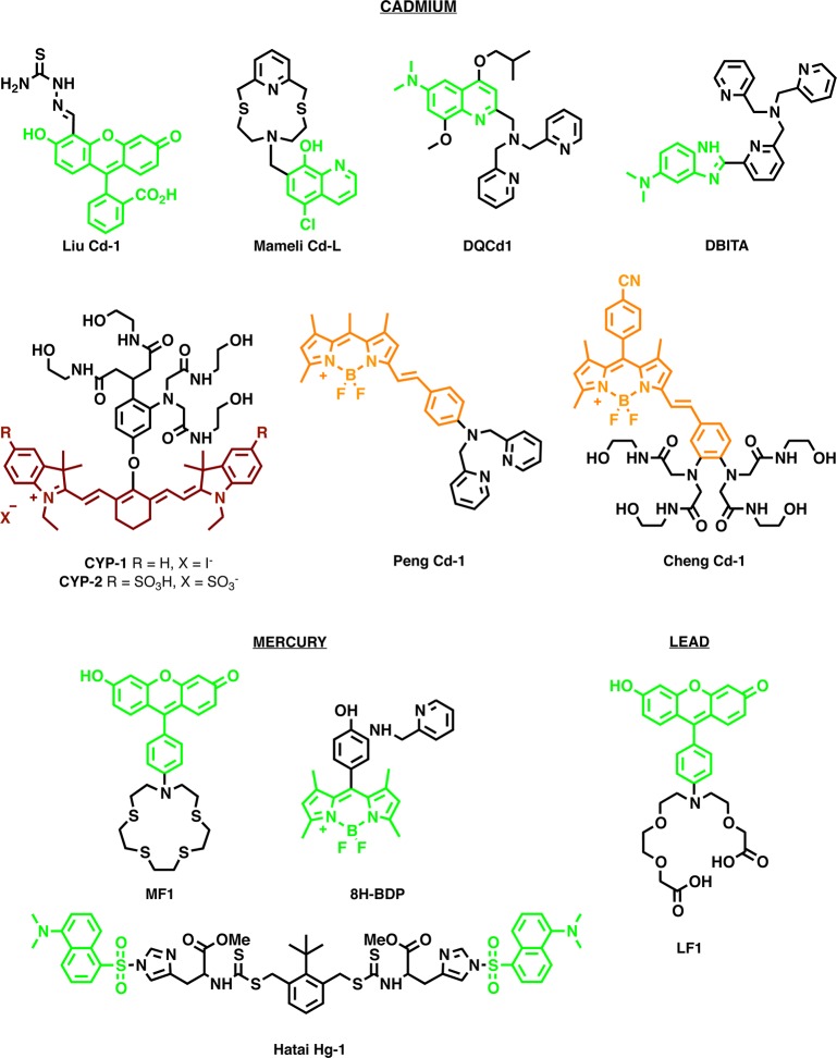
Molecular sensors for the toxic metals Pb2+, Cd2+, and Hg2+.
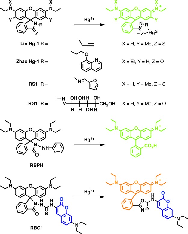
Hg2+ sensor built on a spirolactam
ring-opening platform.
References
-
- Biological Inorganic Chemistry, 1st ed.; Bertini I., Gray H. B., Stiefel E. I., Valentine J. S., Eds.; University Science Books: Sausalito, CA, 2007.
-
- Gladyshev V. N.; Zhang Y. Met. Ions Life Sci. 2013, 12, 529. - PubMed
-
- Cvetkovic A.; Menon A. L.; Thorgersen M. P.; Scott J. W.; Poole F. L. II; Jenney F. E. Jr.; Lancaster W. A.; Praissman J. L.; Shanmukh S.; Vaccaro B. J.; Trauger S. A.; Kalisiak E.; Apon J. V.; Siuzdak G.; Yannone S. M.; Tainer J. A.; Adams M. W. Nature 2010, 466, 779. - PubMed
- Yannone S. M.; Hartung S.; Menon A. L.; Adams M. W.; Tainer J. A. Curr. Opin. Biotechnol. 2012, 23, 89. - PMC - PubMed
-
- Cerchiaro G.; Manieri T. M.; Bertuchi F. R.. Metallomics 2013. - PubMed
Publication types
MeSH terms
Substances
Grants and funding
LinkOut - more resources
Full Text Sources
Other Literature Sources
