. 2014 Mar 3:14:53.
doi: 10.1186/1471-2180-14-53.

Development and antimicrobial susceptibility studies of in vitro monomicrobial and polymicrobial biofilm models with Aspergillus fumigatus and Pseudomonas aeruginosa

Affiliations

Development and antimicrobial susceptibility studies of in vitro monomicrobial and polymicrobial biofilm models with Aspergillus fumigatus and Pseudomonas aeruginosa

Elias K Manavathu et al. BMC Microbiol. .

Abstract

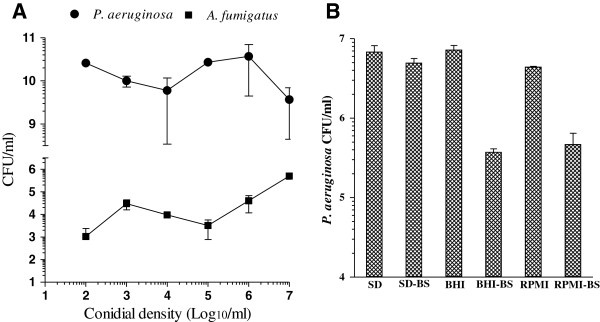
Background: Mixed microbial infections of the respiratory tracts with P. aeruginosa and A. fumigatus capable of producing biofilms are commonly found in cystic fibrosis patients. The primary objective of this study was to develop an in vitro model for P. aeruginosa and A. fumigatus polymicrobial biofilm to study the efficacy of various antimicrobial drugs alone and in combinations against biofilm-embedded cells. Simultaneous static cocultures of P. aeruginosa and sporelings were used for the development of in vitro P. aeruginosa-A. fumigatus polymicrobial biofilm in SD broth in 24-well cell culture plates at 35°C, and the biofilm formation was monitored microscopically and spectrophotometrically. Using P. aeruginosa-A. fumigatus sporelings cocultures we examined the effects of various antimicrobial drugs alone and in combination against polymicrobial biofilm by CFU and tetrazolium reduction assays.

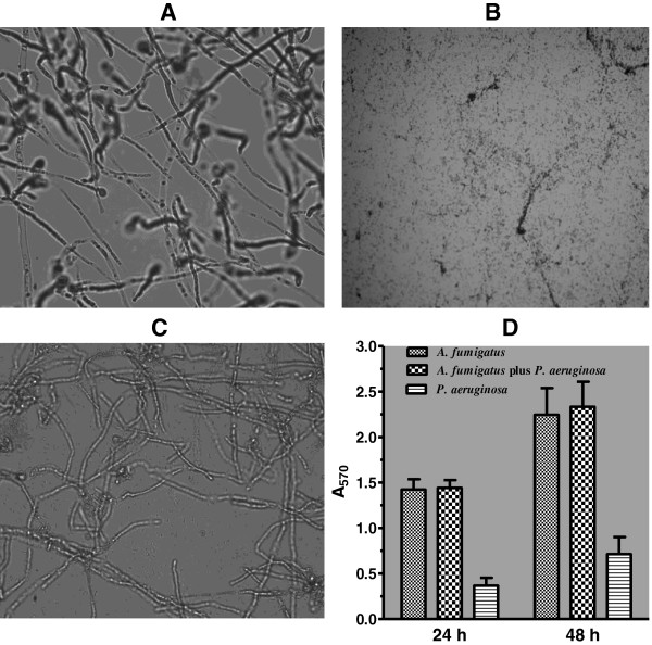
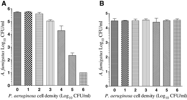
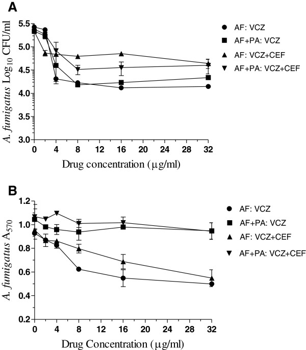
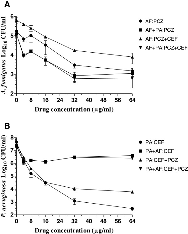
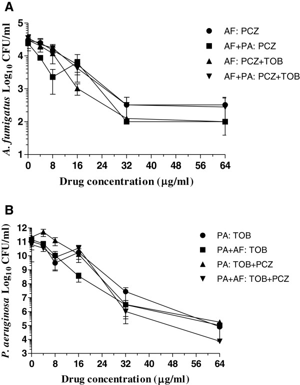
Results: In simultaneous static cocultures P. aeruginosa cells killed A. fumigatus conidia, whereas the bacterial cells showed no substantial fungicidal effect on sporelings grown for 12 h or longer at 35°C. Monospecies cultures of P. aeruginosa produced loosely adhered monomicrobial biofilm and addition of 10% bovine serum to the growth medium inhibited the formation of monomicrobial biofilm by P. aeruginosa whereas it produced tightly adhered polymicrobial biofilm in the presence of A. fumigatus mycelial growth. A. fumigatus produced firmly adherent monomicrobial and polymicrobial biofilms. A comparison of CFU and MTT assays showed that the latter is unsuitable for studying the effectiveness of antimicrobial treatment against polymicrobial biofilm. Tobramycin alone and in combination with posaconazole was highly effective against monomicrobial and polymicrobial biofilms of P. aeruginosa whereas cefepime alone and in combination with posaconazole showed excellent activity against monomicrobial biofilm of P. aeruginosa but was less effective against polymicrobial biofilm. Monomicrobial and polymicrobial biofilms of A. fumigatus showed similar susceptibility to posaconazole with and without the antibacterial drug.

Conclusions: Simultaneous static coculture of A. fumigatus sporelings grown for 12 h or longer was superior to ungerminated conidia with P. aeruginosa for the development of A. fumigatus-P. aeruginosa biofilm. P. aeruginosa-A. fumigatus polymicrobial biofilm shows differential susceptibility to antimicrobial drugs whereas the susceptibility of A. fumigatus to antimicrobial drugs was unchanged.


Figures

Figure 1
Figure 1
Photomicrographic images and quantification of A. fumigatus and P. aeruginosa biofilms.A. Monomicrobial biofilm of AF53470 grown on plastic cover slips for 48 h at 35°C. B. Monomicrobial biofilm of PA56402 grown on plastic cover slips for 48 h at 35°C. C. Polymicrobial biofilm formed in coculture by AF53470 sporelings and PA56402 grown on plastic cover slips for 48 h at 35°C. The biofilms were photographed using a Nikon Microscope Camera System equipped with SPOT image processing computer software [46]. With the SPOT program, each Objective (10× to 100×) of the microscope was calibrated using a stage micrometer as previously described in the SPOT Software User Guide (Chapter 4, pages 76 and 77). The photomicrographs shown in Figure 1 were captured using the 60× Objective providing a total magnification of 600×. D. Quantification of 24-h and 48-h monomicrobial and polymicrobial biofilms of AF53470 and PA56402. The biofilm quantification experiment by crystal violet binding assay was performed two times with eight replications for each group. The data were analyzed by two-way ANOVA and paired Student’s t-test using GraphPad Prism 5.0. The vertical bar on each histogram represents the standard error of the mean for two independent experiments. The laboratory isolates AF36607 and PA27853 also produced similar monomicrobial and polymicrobial biofilms on plastic cover slips and Costar 6-well cell culture plates.
Figure 2
Figure 2
Effects of P. aeruginosa on A. fumigatus conidia (A) and sporelings (B) in cocultures.A. fumigatus conidia (A) and sporelings (B) at a density of 1 × 10⁶ cells/ml were incubated with P. aeruginosa cells ranging from 1 x 101-1 x 10⁶ cells/ml in 1 ml SD broth at 35°C for 24 h. At the end of the incubation the adherent microbial growth containing fungal and bacterial cells were washed 3 times with distilled water (1 ml each) and the viability of the cells was determined by CFU assay. In all mixed cultures the P. aeruginosa CFUs were similar (≈1 × 10¹⁰ CFU/ml). The experiment was performed at two different times with AF53470 and PA56402 using independently prepared conidial suspensions and bacterial cultures, and one time with AF36607 and PA27853. Similar results were obtained for the clinical and the laboratory isolates. The vertical bar on each data point represents the standard error of the mean for two independent experiments with AF53470 and PA56402. The data were analyzed by one way ANOVA with Dunnett multiple comparison test where the control was compared with each of the experimental group using GraphPad Prism 5.0.
Figure 3
Figure 3
Effects of cell density and growth medium on biofilm formation. A. Effect of conidial density on A. fumigatus-P. aeruginosa polymicrobial biofilm formation. One ml aliquots of AF53470 conidial suspension containing 1 × 10² - 1 × 10⁷ conidia/ml were incubated in 24-well cell culture plates in duplicates at 35°C in SD broth for 18 h, washed and then inoculated with 1 × 10⁶ PA56402 cells in 1 ml SD broth and further incubated for 24 h for the development of A. fumigatus-P. aeruginosa polymicrobial biofilm. The biofilm was washed and the embedded cells were resuspended in 1 ml sterile water and assayed for A. fumigatus and P. aeruginosa by CFU counts. The experiment was performed at two different times using independently prepared conidial suspensions and bacterial cultures and the vertical bar on each data point on the graph represents the standard error of the mean. B. P. aeruginosa monomicrobial biofilm formation in various growth media with and without bovine serum. One ml aliquots of growth media containing 1 × 10⁶P. aeruginosa cells were incubated in quadruplicates in 24-well cell culture plates with and without 10% bovine serum for 24 h at 35°C for biofilm formation. The adherent monomicrobial biofilm was washed (3 times), resuspended in 1 ml sterile distilled water and the biofilm growth was assessed by CFU assay. The experiment was performed two different times with PA56402 using independently prepared bacterial cultures, and one time with PA27853. Both sets of isolates provided similar results. The data were analyzed by paired Student’s t test using GraphPad prism 5.0. The vertical bar on each histogram denotes standard error of the mean for two independent experiments using PA56402. Legends: SD, Sabouraud’s dextrose broth; SD-BS, Sabouraud’s dextrose broth with 10% bovine serum; BHI, Brain Heart Infusion broth; BHI-BS Brain Heart Infusion broth with 10% bovine serum; RPMI, RPMI640; RPMI-BS, RPMI1640 with 10% bovine serum.
Figure 4
Figure 4
Effects of voriconazole alone and in combination with cefepime against A. fumigatus monomicrobial and A. fumigatus-P. aeruginosa polymicrobial biofilms as determined by CFU (A) and MTT (B) assays. The biofilms were developed in 24-well cell culture plates and the effectiveness of antimicrobial drug(s) treatment was assessed by the reduction of CFUs and A570 values. Each experiment was performed two different times with the clinical isolates AF53470 and PA56402 using independently prepared conidial suspensions and bacterial cultures, and one time with the laboratory isolates AF36607 and PA27853. Similar results were obtained for both set of isolates. The data were analyzed by two-way ANOVA with Bonferroni post test analysis by comparing each treatment group to the other for statistical significance using Graphpad Prism 5.0. The vertical bar on each data point denotes standard error of the mean for two experiments performed with AF53470 and PA56402. Legends: AF, A. fumigatus monomicrobial biofilm; AF + PA, A. fumigatus-P. aeruginosa polymicrobial biofilm; VCZ, voriconazole; CEF, cefepime.
Figure 5
Figure 5
Biofilm inhibition by posaconazole and cefepime. A. Effects of posaconazole alone and in combination with cefepime against A. fumigatus monomicrobial and A. fumigatus-P. aeruginosa polymicrobial biofilms. B. Effects of cefepime alone and in combination with posaconazole against P. aeruginosa monomicrobial and P. aeruginosa-A. fumigatus polymicrobial biofilms. Each experiment was performed two different times with the clinical isolates AF53470 and PA57402 using independently prepared conidial suspensions and bacterial cultures, and one time with the laboratory isolates AF36607 and PA27853. Both clinical and laboratory isolates provided similar results. The data were analyzed by one-way and two-way ANOVA with Bonferroni’s multiple comparison test where each set of data is compared with all the other sets of data as well as by paired two-tailed Student’s t-test using Graphpad Prism 5.0. The vertical bar on each data point denotes standard error of the mean for two independent experiments performed with the clinical isolates. Legends: AF, A. fumigatus monomicrobial biofilm; PA, P. aeruginosa monomicrobial biofilm; PA + AF and AF + PA, polymicrobial biofilm; CEF, cefepime; PCZ, posaconazole.
Figure 6
Figure 6
Biofilm inhibition by posaconazole and tobramycin. A. Effects of posaconazole alone and in combination with tobramycin against A. fumigatus monomicrobial and A. fumigatus-P. aeruginosa polymicrobial biofilms. B. Effects of tobramycin alone and in combination with posaconazole against P. aeruginosa monomicrobial and P. aeruginosa-A. fumigatus polymicrobial biofilms. Each experiment was performed two different times with the clinical isolates AF53470 and PA57402 using independently prepared conidial suspensions and bacterial cultures, and one time with the laboratory isolates AF36607 and PA27853. Both clinical and laboratory isolates provided similar results. The data were analyzed by one-way and two-way ANOVA with Bonferroni’s multiple comparison test where each set of data is compared with all the other sets of data as well as by paired two-tailed Student’s t-test using Graphpad Prism 5.0. The vertical bar on each data point denotes standard error of the mean for two independent experiments performed with the clinical isolates. Legends: AF, A. fumigatus monomicrobial biofilm; PA, P. aeruginosa monomicrobial biofilm; AF + PA and PA + AF, polymicrobial biofilm; PCZ, posaconazole; TOB, tobramycin.

Similar articles

Cited by

References

    1. Zwielehner J, Lassl C, Hippe B, Pointner A, Switzeny OJ, Remely M, Kitzweger E, Ruckser R, Haslberger AG. Changes in human fecal microbiota due to chemotherapy analyzed by TaqMan-PCR, 454 sequencing and PCR-DGGE fingerprinting. PLoS One. 2011;6:e28654. doi: 10.1371/journal.pone.0028654. - DOI - PMC - PubMed
    1. Charlson ES, Diamond JM, Bittinger K, Fitzgerald AS, Yadav A, Haas AR, Bushman FD, Collman RG. Lung-enriched organisms and aberrant bacterial and fungal respiratory microbiota after lung transplant. Am J Respir Crit Care Med. 2012;186:536–545. doi: 10.1164/rccm.201204-0693OC. - DOI - PMC - PubMed
    1. Iwai S, Fei M, Huang D, Fong S, Subramanian A, Grieco K, Lynch SV, Huang L. Oral and airway microbiota in HIV-infected pneumonia patients. J Clin Microbiol. 2012;50:2995–3002. doi: 10.1128/JCM.00278-12. - DOI - PMC - PubMed
    1. Rolston KV, Bodey GP, Safdar A. Polymicrobial infection in patients with cancer: an underappreciated and underreported entity. Clin Infect Dis. 2007;45:228–233. doi: 10.1086/518873. - DOI - PubMed
    1. Duggal R, Rajwanshi A, Gupta N, Lal A, Singhal M. Polymicrobial lung infection in postrenal transplant recipient diagnosed by fine-needle aspiration cytology. Diagn Cytopathol. 2010;38:294–296. - PubMed

Publication types

MeSH terms