. 2014 May-Jun;152(1-2):77-89.
doi: 10.1016/j.clim.2014.02.010. Epub 2014 Mar 1.

Identifying functional anti-Staphylococcus aureus antibodies by sequencing antibody repertoires of patient plasmablasts

Affiliations

Identifying functional anti-Staphylococcus aureus antibodies by sequencing antibody repertoires of patient plasmablasts

Daniel R Lu et al. Clin Immunol. 2014 May-Jun.

Abstract

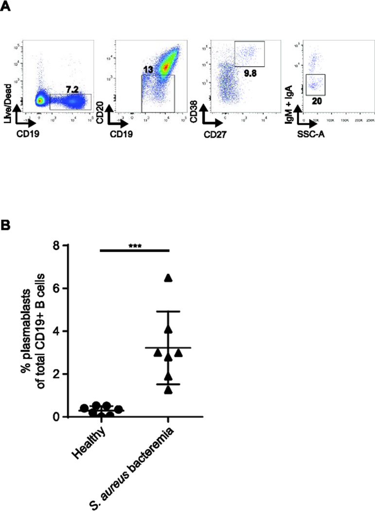
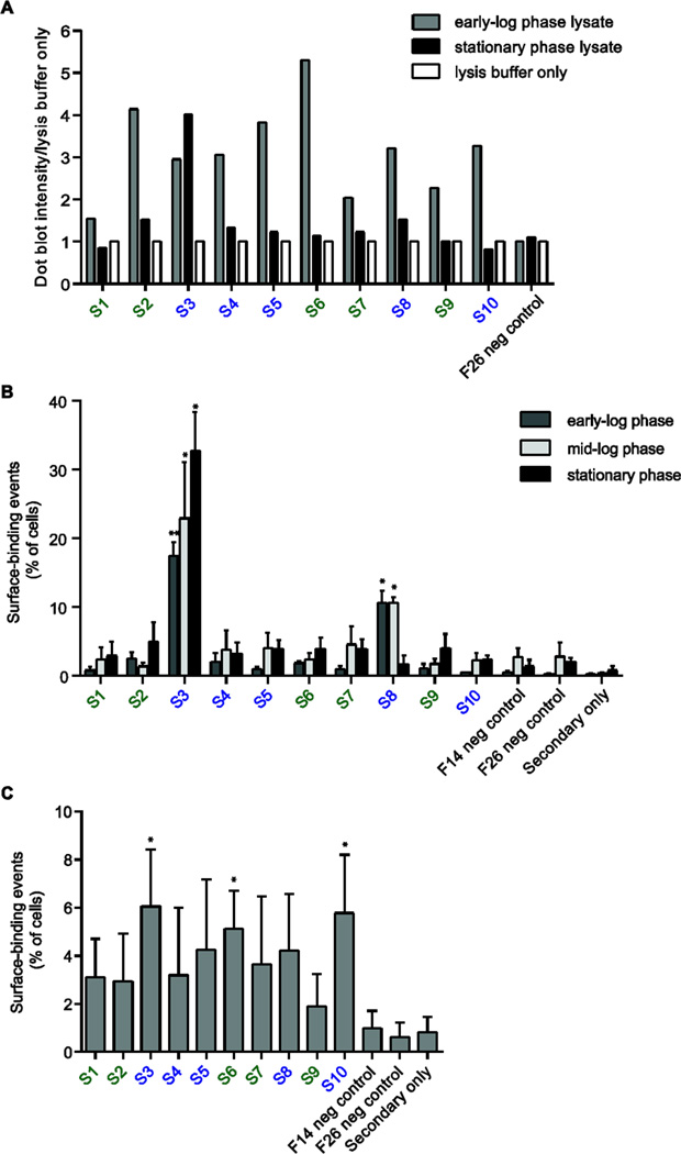
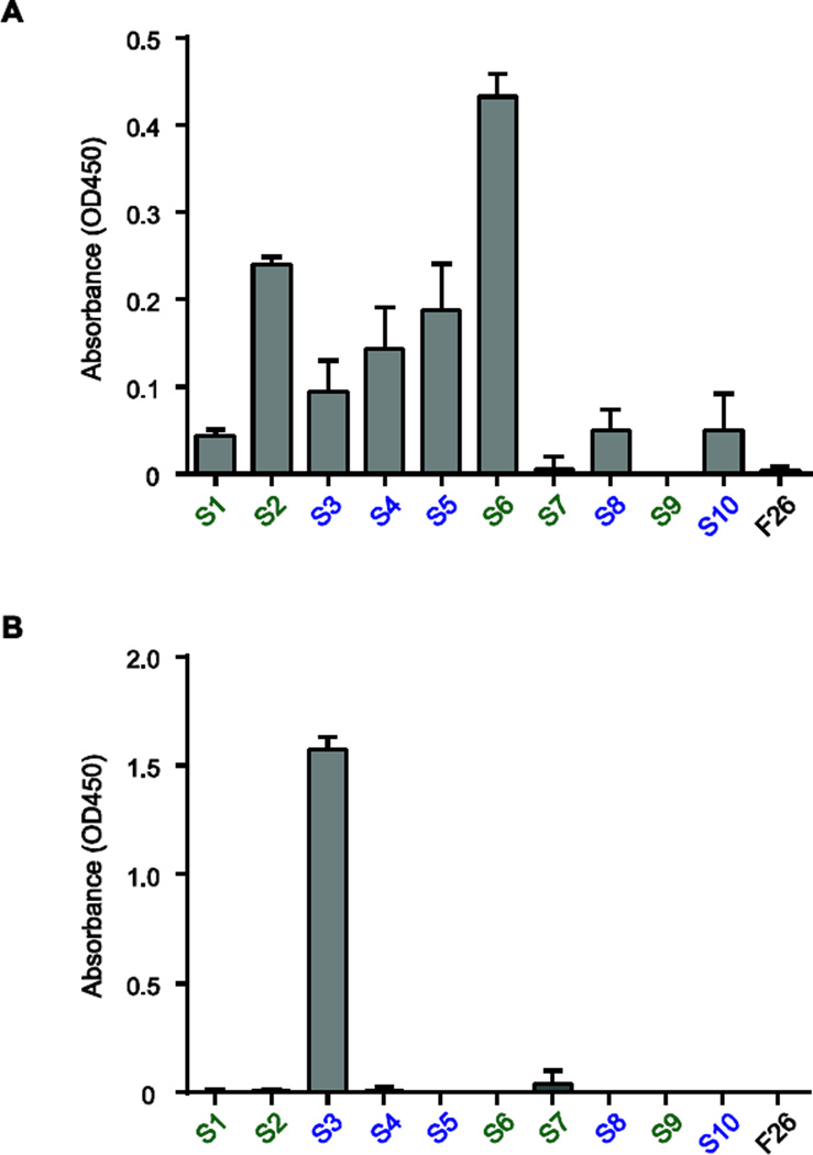
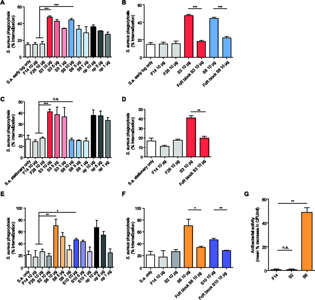
Infection by Staphylococcus aureus is on the rise, and there is a need for a better understanding of host immune responses that combat S. aureus. Here we use DNA barcoding to enable deep sequencing of the paired heavy- and light-chain immunoglobulin genes expressed by individual plasmablasts derived from S. aureus-infected humans. Bioinformatic analysis of the antibody repertoires revealed clonal families of heavy-chain sequences and enabled rational selection of antibodies for recombinant expression. Of the ten recombinant antibodies produced, seven bound to S. aureus, of which four promoted opsonophagocytosis of S. aureus. Five of the antibodies bound to known S. aureus cell-surface antigens, including fibronectin-binding protein A. Fibronectin-binding protein A-specific antibodies were isolated from two independent S. aureus-infected patients and mediated neutrophil killing of S. aureus in in vitro assays. Thus, our DNA barcoding approach enabled efficient identification of antibodies involved in protective host antibody responses against S. aureus.

Keywords: Antibody; Infection; Staphylococcus aureus.


Figures

Figure 1
Figure 1
Plasmablasts are elevated during acute S. aureus bacteremia. (A) Fluorescenceactivated gating strategy for single-cell sorting IgG-secreting plasmablasts from PBMCs of S. aureus-infected individuals. Plasmablasts were gated on CD19+/intCD20CD27++CD38++IgAIgM live B cells. (B)
Figure 2
Figure 2
Heavy-chain dendrograms of the plasmablast antibody repertoires of S. aureus-bacteremic individuals and shared HC V-J analysis. Each peripheral node depicts a sequenced VH region with a unique plate and well-ID barcode combination. Plasmablast antibody repertoires of two individuals with S. aureus bacteremia: (A) patient 1 and (B) patient 2. Red lines indicates the IGHV families with greater than 5 recovered full-length VH sequences, from which analysis of shared IGHJ was performed. Pie charts of the IGHJ gene usage for each IGHV family are displayed adjacent to the branch representing each IGHV. For presentation, red bolded lines indicate the IGHV families where a prominent IGHJ gene was used. S1, S2, S3, S4, S5, S6, S7, S8, S9, and S10 denote the recombinant antibodies that were selected for recombinant expression. Circles denote antibodies that bind S. aureus, and squares denote antibodies that do not. Recombinant antibodies from shared gene families are denoted in blue, and antibodies not sharing gene rearrangements are denoted in green.
Figure 3
Figure 3
Screening the binding activity of recombinant antibodies generated from the plasmablasts of S. aureus-infected individuals. (A) Dot-blot analysis of antibody binding to lysates prepared from S. aureus Wood46 at early-log and stationary growth phases. Data are dot-blot densitometry values shown as a fold increase relative to control (lysis buffer only). (B,C) Flow cytometric detection of antibody binding to the surface of S. aureus Wood46 at different growth phases in LB (B) or in the presence of NIH/3T3 fibroblasts (C). Recombinant antibodies from shared gene families are denoted in blue, and antibodies not sharing gene rearrangements are denoted in green. Influenza-specific antibodies F14 and F26 were used as isotype controls. Data in (B) and (C) are shown as the mean ± s.e.m. of triplicates and are representative of three or more independent experiments *P < 0.05 by two-tailed unpaired t-test, comparing binding of S. aureus recombinant antibodies to that of the isotype control antibodies.
Figure 4
Figure 4
Identification of protein antigens targeted by plasmablast-derived recombinant antibodies. (A) Screen of recombinant antibody binding to a S. aureus USA300 proteome microarray. Recombinant antibodies from shared gene families are denoted in blue, and antibodies not sharing gene rearrangements are denoted in green. Measurement of antibody binding ranged from 5000 units (low-affinity) to 20,000 units (high-affinity). Mean fluorescence intensity is displayed and the scale bar represents the range of fluorescence units measured. Only antigens with a binding intensity greater than two standard deviations of signal above background are shown. (B) Schematic representation of the recombinant full-length and truncated N-terminal and C-terminal fragments of FnBPA with domains annotated; numbers indicate the amino-acid residue position. (C) Antibody-binding assay plotting S6 and S10 binding against recombinant full-length, truncated N-terminal, and truncated C-terminal fragments of FnBPA.
Figure 5
Figure 5
Identification of S. aureus cell wall components targeted by plasmablast-derived recombinant antibodies. (A) ELISA of recombinant antibody binding to S. aureus peptidoglycan. (B) ELISA of recombinant antibody binding to S. aureus lipoteichoic acid. Recombinant antibodies from shared gene families are denoted in blue, and antibodies not sharing gene rearrangements are denoted in green. Influenza-specific antibody F26 was used as an isotype control. ELISAs were performed in triplicate and are presented as mean values of all replicates ± s.e.m.
Figure 6
Figure 6
Opsonophagocytic and bactericidal capability of recombinant antibodies generated from S. aureus-infected individuals. (A,C,E) Flow cytometric analysis of antibody-mediated phagocytosis. CFSE-labeled S. aureus Wood46 fixed in early-log growth phase (A), fixed in stationary growth phase (C), or grown with NIH/3T3 fibroblasts (E), were incubated with undifferentiated THP1 monocyte-like cells and 1 µg/ml, 5 µg/ml, or 10 µg/ml of recombinant antibody. (B,D,F) Flow cytometric analysis of FcR dependence of antibody-mediated phagocytosis. Early-log phase (B), stationary phase (D), and NIH/3T3-co-cultured (F), antibodyopsonized, CFSE-labeled S. aureus Wood46 were incubated with undifferentiated THP1 cells pre-treated with FcR-blocking human IgG. (G) Evaluation of the antibodies’ antibacterial activity. Live S. aureus Wood46 were incubated with antibodies and activated HL-60 neutrophils. Bacteria were plated following HL-60 lysis, and mean percent decrease in CFU was measured. Flu-specific antibodies F14 and F26 were used as isotype controls. op = anti-S. aureus polyclonal rabbit IgG antibodies. Data are from three experiments performed on separate days and are presented as mean values of all replicates ± s.e.m. Statistical significance was determined by using two-tailed unpaired t-tests; n.s., not significant (P > 0.05); *P < 0.05; **P < 0.01; ***P < 0.001.

References

    1. Talan DA, et al. Comparison of Staphylococcus aureus from skin and soft-tissue infections in US emergency department patients 2004 and 2008. Clin Infect Dis. 2011;53(2):144–149. - PubMed
    1. Klein E, Smith DL, Laxminarayan R. Hospitalizations and deaths caused by methicillinresistant Staphylococcus aureus, United States 1999–2005. Emerg Infect Dis. 2007;13(12):1840–1846. - PMC - PubMed
    1. Klevens RM, et al. Invasive methicillin-resistant Staphylococcus aureus infections in the United States. JAMA. 2007;298(15):1763–1771. - PubMed
    1. Proctor RA. Challenges for a universal Staphylococcus aureus vaccine. Clin Infect Dis. 2012;54(8):1179–1186. - PubMed
    1. Holtfreter S, Kolata J, Broker BM. Towards the immune proteome of Staphylococcus aureus - The anti-S. aureus antibody response. Int J Med Microbiol. 2010;300(2–3):176–192. - PubMed

Publication types

MeSH terms

Substances