. 2014 Mar 1;28(5):432-7.
doi: 10.1101/gad.233676.113.

A temporal requirement for Hippo signaling in mammary gland differentiation, growth, and tumorigenesis

Affiliations

A temporal requirement for Hippo signaling in mammary gland differentiation, growth, and tumorigenesis

Qian Chen et al. Genes Dev. .

Abstract

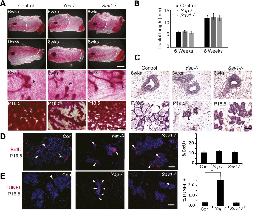
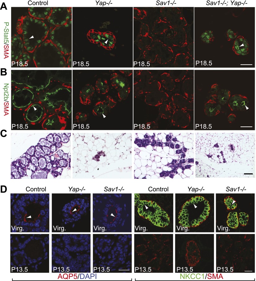
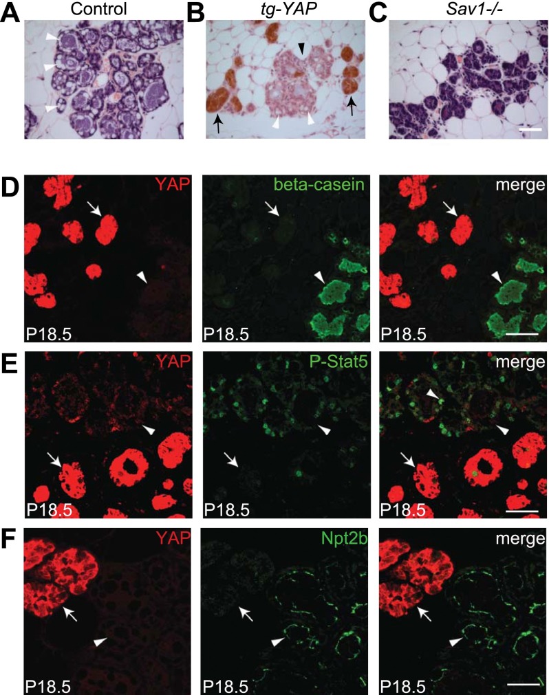
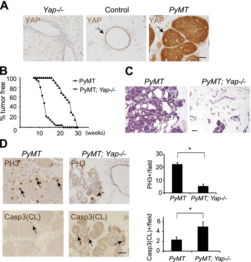
Despite recent progress, the physiological role of Hippo signaling in mammary gland development and tumorigenesis remains poorly understood. Here we show that the Hippo pathway is functionally dispensable in virgin mammary glands but specifically required during pregnancy. In contrast to many other tissues, hyperactivation of YAP in mammary epithelia does not induce hyperplasia but leads to defects in terminal differentiation. Interestingly, loss of YAP causes no obvious defects in virgin mammary glands but potently suppresses oncogene-induced mammary tumors. The selective requirement for YAP in oncogenic growth highlights the potential of YAP inhibitors as molecular targeted therapies against breast cancers.

Keywords: Hippo signaling; YAP; differentiation; mammary gland; proliferation; tumorigenesis.


Figures

Figure 1.
Figure 1.
Impaired development in Yap-deficient and Sav1-deficient mammary glands. (A) Whole-mount staining of control (MMTV-Cre only), Yap-deficient, and Sav1-deficient mammary glands. (Top two rows) Low-magnification view of 6- and 8-wk virgin glands. Note the similar appearance and ductal invasion in each genotype. Bar, 5mm. (Bottom two rows) High-magnification view of 6-wk virgin and P18.5 glands. Note the normal appearance of terminal end buds of Yap−/− or Sav1−/− virgin glands. Also note the greatly reduced lobuloalveolar structures of Yap-deficient glands and the tightly packed and undistended alveoli of Sav1-deficient glands in P18.5 glands. Bar, 500 μm. (B) Quantification of ductal invasion in 6- and 8-wk virgin glands, measured as the distance between the distal end of the lymph node and the distal end of the longest duct. Data are mean ± SEM; n = 3. P > 0.05, t-test. (C) H&E staining of control (MMTV-Cre only), Yap-deficient, and Sav1-deficient mammary glands from 6-wk virgin and P18.5 glands. Note the normal appearance of terminal end buds in Yap−/− or Sav1−/− virgin glands. In P18.5 animals, fewer ducts and lobuloalveoli were present in Yap-deficient glands, and the remaining lobuloalveoli were significantly smaller and had fewer cells per alveolus compared with control. Sav1-deficient lobuloalveoli were undistended but contained a normal number of cells per alveolus. Also note the presence of lipid droplets (arrowheads) in control and Yap mutant glands but not in Sav1 mutant glands at P18.5. Bar , 50 μm. (D) BrdU analysis of P16.5 glands. Arrowheads mark selected BrdU-positive cells. Note the similar number of BrdU-positive cells in each genotype. The graph shows mean ± SEM, n = 5. P > 0.05, t-test. Bar, 100 μm. (E) TUNEL analysis. Arrowheads mark examples of TUNEL-positive cells. Note the significant increase of TUNEL-positive cells in Yap-deficient, but not Sav1-deficient, mammary glands. The graph shows mean ± SEM; n = 5. (*) P < 0.05, t-test. Bar, 100 μm.
Figure 2.
Figure 2.
Loss of Sav1 leads to defects in terminal differentiation of mammary glands. (A–C) P18.5 mammary glands were analyzed for P-Stat5 (green) and smooth muscle actin (SMA, red) (A), Npt2b (green) and SMA (red) (B), and H&E staining (C). Note the presence of P-Stat5 and Npt2b staining in Yap−/− and Sav1−/− Yap−/− glands and its absence in Sav1−/− glands. Bars, 20 μm. (D) Control (MMTV-Cre only), Yap-deficient, and Sav1-deficient mammary glands at P13.5 were stained for AQP5 (red) plus DAPI (blue) or NKCC1 (green) plus SMA (red). Note the presence of AQP5 and NKCC1 signals (arrowheads) in virgin but not P13.5 glands in all genotypes. Bar, 20 μm.
Figure 3.
Figure 3.
YAP overexpression causes terminal differentiation defects similar to loss of Sav1. (A) H&E staining of a wild-type P18.5 mammary gland. Note the distended alveolar morphology and the cytoplasmic accumulation of lipid droplets (arrowheads). (B) A P18.5 MMTV-rtTA; TRE-YAP mammary gland stained for YAP (brown) and counterstained by H&E. Note the mosaic pattern of YAP transgene induction, showing areas with (arrows) and without (arrowhead) YAP overexpression. Also note the nondistended morphology of YAP-overexpressing alveoli (arrow), while the neighboring nonoverexpressing alveoli (black arrowhead) showed distended morphology and cytoplasmic lipid droplets (white arrowheads). (C) H&E staining of a P18.5 Sav1 mammary gland. Note the undistended morphology and the absence of cytoplasmic lipid droplets. Bar, 50 μm. (D–F) P18.5 MMTV-rtTA; TRE-YAP transgenic mammary glands stained for YAP (red) and β-casein (D), P-Stat5 (E), or Npt2b (F). Note the absence of these differentiation markers (green) in YAP-overexpressing alveoli (arrow) and the presence of these markers in nonoverexpressing alveoli (arrowhead). Bar, 20 μm.
Figure 4.
Figure 4.
Loss of YAP suppresses PyMT-induced tumor growth. (A) Twelve-week-old Yap-deficient, control, and PyMT virgin glands were stained for YAP. Note the absence of YAP staining in the Yap-deficient mammary epithelia and elevated YAP expression in the PyMT mammary tumors (arrows). Bar, 50 μm. (B) Fraction of mammary pads free of palpable tumors in the PyMT (black diamond; n = 33) and Yap-deficient PyMT (black triangle; n = 20) virgin mice. (C) H&E staining of 15-wk-old PyMT and Yap-deficient PyMT virgin glands. Note the significant suppression of mammary ductal hyperplasia in the Yap-deficient PyMT gland. Bar, 200 μm. (D) Analysis of cell proliferation and cell death in 15-wk-old PyMT and Yap-deficient PyMT virgin glands. Quantification is shown in the graphs. Note decreased cell proliferation and increased apoptosis in the Yap-deficient PyMT mice. (*) P < 0.05, t-test. Bar, 50 μm.
Figure 5.
Figure 5.
VP suppresses the growth of human breast cancer cell lines. Cells were seeded in 96-well plates with 0, 1, 3, or 10 µM VP. Cell number was measured at 0–5 d. Note the differential sensitivity of the cell lines to VP.

Similar articles

Cited by

References

    1. Brisken C, Rajaram RD 2006. Alveolar and lactogenic differentiation. J Mammary Gland Biol Neoplasia 11: 239–248 - PubMed
    1. Cai J, Zhang N, Zheng Y, de Wilde RF, Maitra A, Pan D 2010. The Hippo signaling pathway restricts the oncogenic potential of an intestinal regeneration program. Genes Dev 24: 2383–2388 - PMC - PubMed
    1. Chan SW, Lim CJ, Guo K, Ng CP, Lee I, Hunziker W, Zeng Q, Hong W 2008. A role for TAZ in migration, invasion, and tumorigenesis of breast cancer cells. Cancer Res 68: 2592–2598 - PubMed
    1. Chen D, Sun Y, Wei Y, Zhang P, Rezaeian AH, Teruya-Feldstein J, Gupta S, Liang H, Lin HK, Hung MC, et al. 2012. LIFR is a breast cancer metastasis suppressor upstream of the Hippo–YAP pathway and a prognostic marker. Nat Med 18: 1511–1517 - PMC - PubMed
    1. Cordenonsi M, Zanconato F, Azzolin L, Forcato M, Rosato A, Frasson C, Inui M, Montagner M, Parenti AR, Poletti A, et al. 2011. The hippo transducer TAZ confers cancer stem cell-related traits on breast cancer cells. Cell 147: 759–772 - PubMed

Publication types

Substances