. 2014 Apr;124(4):1622-35.
doi: 10.1172/JCI71386. Epub 2014 Mar 3.

Fibrotic extracellular matrix activates a profibrotic positive feedback loop

Fibrotic extracellular matrix activates a profibrotic positive feedback loop

Matthew W Parker et al. J Clin Invest. 2014 Apr.

Abstract

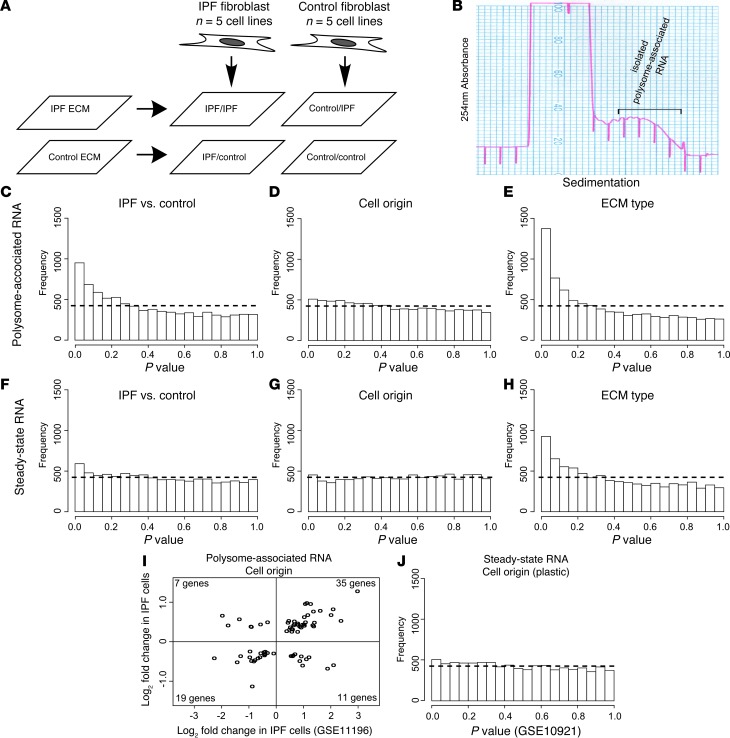
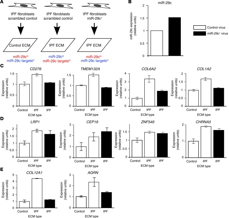
Pathological remodeling of the extracellular matrix (ECM) by fibroblasts leads to organ failure. Development of idiopathic pulmonary fibrosis (IPF) is characterized by a progressive fibrotic scarring in the lung that ultimately leads to asphyxiation; however, the cascade of events that promote IPF are not well defined. Here, we examined how the interplay between the ECM and fibroblasts affects both the transcriptome and translatome by culturing primary fibroblasts generated from IPF patient lung tissue or nonfibrotic lung tissue on decellularized lung ECM from either IPF or control patients. Surprisingly, the origin of the ECM had a greater impact on gene expression than did cell origin, and differences in translational control were more prominent than alterations in transcriptional regulation. Strikingly, genes that were translationally activated by IPF-derived ECM were enriched for those encoding ECM proteins detected in IPF tissue. We determined that genes encoding IPF-associated ECM proteins are targets for miR-29, which was downregulated in fibroblasts grown on IPF-derived ECM, and baseline expression of ECM targets could be restored by overexpression of miR-29. Our data support a model in which fibroblasts are activated to pathologically remodel the ECM in IPF via a positive feedback loop between fibroblasts and aberrant ECM. Interrupting this loop may be a strategy for IPF treatment.


Figures

Figure 1
Figure 1. Pathological gene expression in fibrotic fibroblasts is primarily governed by diseased ECM.
(A) Experimental design. IPF or control fibroblasts were cultured on IPF or control ECM using a 2 × 2 experimental design. (B) Isolation of polysome-associated RNA. Sample polysome tracing shows absorbance at 254 nm across the sucrose gradient. Transcripts that are translated are enriched among polysome-associated RNA. The portion of the gradient collected to measure polysome-associated RNA levels is indicated. (CH) Histograms of gene-by-gene P values for different biological comparisons. Dashed lines represent theoretical null distributions. (CE) Comparison of polysome-associated RNA levels of (C) IPF fibroblasts seeded on IPF ECM and control fibroblasts seeded on control ECM, (D) IPF fibroblasts and control fibroblasts (independent of ECM type), and (E) IPF and control ECM (independent of cell origin). (FH) Same comparisons as in AC using steady-state RNA data. (I) Genes that showed cell origin modulation in polysome-associated RNA (P < 0.05) in both our current study and in a previous study (microarray data obtained from GSE11196) were collected. Plotted are the fold changes in the previous study (x axis) and in our current dataset (y axis). Genes upregulated in one study are more likely to be upregulated in the other (κ = 0.476, 95% CI = [0.269, 0.682]). (J) Histogram of P values for comparison of RNA obtained from control and IPF cells grown under standard culture conditions (microarray data obtained from GEO GSE10921).
Figure 2
Figure 2. Diseased ECM predominantly affects gene expression by modulating translation.
(A and B) Cumulative FDR probability distributions for three control levels: polysome-associated RNA, steady-state RNA, and translation (ANOTA-corrected polysome-associated RNA). Each line indicates the fraction of genes (y axis) that passed a given FDR threshold (x axis) for each control level. (A) Comparison between IPF and control ECM. (B) Comparison between IPF and control fibroblasts. (C) Density plot of gene-by-gene fold changes from the ECM origin comparison. (D) Density plot of gene-by-gene variances in comparisons of ECM origin. (E) Heatmap of genes that were upregulated (yellow) or downregulated (blue) at the steady-state RNA or translation level (FDR < 0.3). Pr, probability. Abs, absolute value; MSS, mean sum of squares.
Figure 3
Figure 3. Diseased ECM coordinately activates the translation of ECM region genes.
(AD) Modulation of genes in the ECM region gene ontology. Upper panels show volcano plots of ECM and cell effects at the translation and steady-state RNA levels. The number of genes up- or downregulated (P < 0.05) is indicated. Lower panels show cumulative P value probability distributions from the same comparisons. Dotted line indicates the theoretical null distribution. Solid black line indicates the distribution for all genes. Red line represents the distribution for those genes in the ECM region gene ontology.
Figure 4
Figure 4. Converging and independent modulation of ECM gene translation is dependent upon ECM and cell origins.
(A) Translation (ANOTA-corrected) and steady-state RNA profiles of genes in the ECM region gene ontology that were differentially expressed (P < 0.05) in any comparison. Values are log10P values (yellow denotes upregulation in IPF, blue denotes downregulation). (B) Close-up of the translation profile. Genes are divided into three categories: cell-regulated, ECM-regulated, and coregulated. Density plots of the absolute fold changes induced by each biological variable are shown. Selected gene ontologies that are overrepresented (P < 0.01, calculated using Fisher’s exact test) are shown (see Supplemental Table 2 for the complete list). There were no significantly overrepresented gene ontologies in the cell-regulated group. (C) Same analysis for the steady-state RNA profile.
Figure 5
Figure 5. A positive feedback loop between diseased ECM and the fibroblast involves modulation of miR-29 expression.
(A) Translation profile of genes whose protein products are present in IPF lung ECM. Shown are –log10P values from the cell origin and ECM origin comparison (yellow denotes upregulation in IPF, blue denotes downregulation). miR-29 targets are designated by solid bars above the heatmap. (B and C) Cumulative P value probability distributions for (B) ECM origin and (C) cell origin are shown. Dotted line represents the theoretical null distribution. Black line represents all genes except ECM region genes. Red line represents all ECM genes except those detected in the IPF lung (KS P value for comparison with “All genes”). Blue line represents all genes from the IPF lung except miRNA-29 targets (KS P value for comparison with “ECM genes”). Yellow line represents all IPF-detected miR-29 targets (KS P value for comparison with “IPF-detected proteins”). (D and E) Levels of miR-29 species were quantified using qPCR. In all comparisons, the arbitrary units were normalized to the control level. The levels of miR-29c between control and IPF ECM were significantly altered (P = 0.031). It should be noted that the ECM comparison is paired so the single error bar represents the standard error between the paired differences.
Figure 6
Figure 6. Overexpression of miR-29c abrogates pathological gene expression on IPF ECM.
(A) Experimental design of miR-29c function study. (B) Relative expression of miR-29c in IPF cells treated with miR-29c+ virus or with scrambled control virus. (C) Relative expression of four genes containing miR-29 targets quantified using qPCR from polysome-associated RNA. White bars represent samples treated with control virus. Black bars represent samples treated with miR-29+ virus. Data represent the mean ± SEM (three technical replicates). (D) Relative expression of four control genes. Controls were identified as genes that microarray indicated were upregulated by IPF but that did not contain miR-29 targets. (E) Relative expression of four ECM genes. ECM genes were identified as being upregulated by IPF but did not contain miR-29 targets.
Figure 7
Figure 7. Positive feedback between the fibrotic ECM and the fibroblast amplifies the fibrotic phenotype.
The IPF ECM induces translation of the genes that comprise the IPF ECM. This induces a positive feedback loop that amplifies ECM gene expression and spreads the fibrosis.

References

    1. Halliday NL, Tomasek JJ. Mechanical properties of the extracellular matrix influence fibronectin fibril assembly in vitro. Exp Cell Res. 1995;217(1):109–117. doi: 10.1006/excr.1995.1069. - DOI - PubMed
    1. Pelham RJ, Jr, Wang Y. Cell locomotion and focal adhesions are regulated by the mechanical properties of the substrate. Biol Bull. 1998;194(3):348–350. doi: 10.2307/1543109. - DOI - PubMed
    1. Rhudy RW, McPherson JM. Influence of the extracellular matrix on the proliferative response of human skin fibroblasts to serum and purified platelet-derived growth factor. J Cell Physiol. 1988;137(1):185–191. doi: 10.1002/jcp.1041370123. - DOI - PubMed
    1. Schor SL. Cell proliferation and migration on collagen substrata in vitro. J Cell Sci. 1980;41(1):159–175. - PubMed
    1. Bhowmick NA, Neilson EG, Moses HL. Stromal fibroblasts in cancer initiation and progression. Nature. 2004;432(7015):332–337. doi: 10.1038/nature03096. - DOI - PMC - PubMed

Publication types

MeSH terms