. 2014 May;58(5):2754-62.
doi: 10.1128/AAC.02565-13. Epub 2014 Mar 3.

Mechanism of Escherichia coli resistance to Pyrrhocoricin

Affiliations

Mechanism of Escherichia coli resistance to Pyrrhocoricin

Shalini Narayanan et al. Antimicrob Agents Chemother. 2014 May.

Abstract

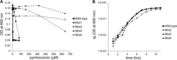
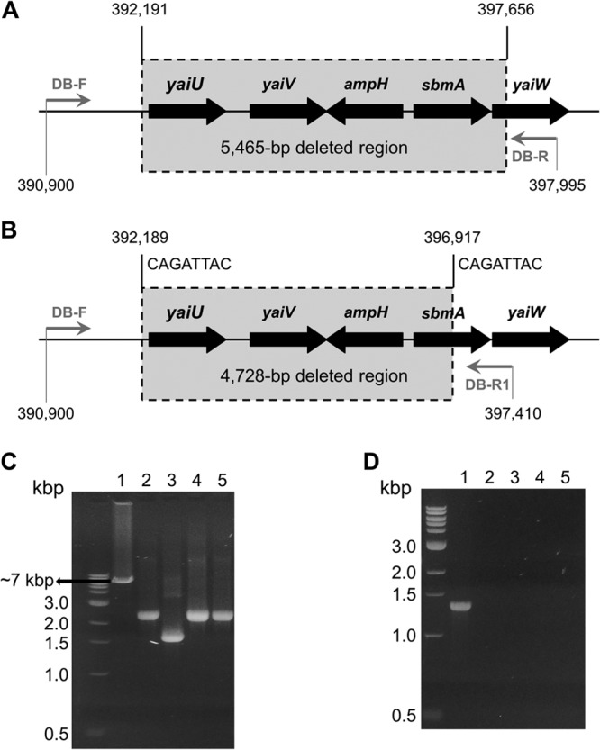
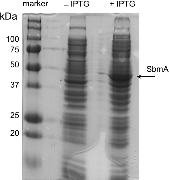
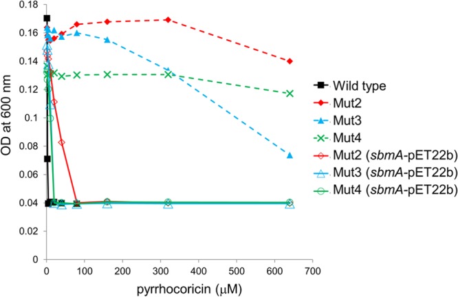
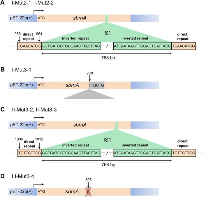
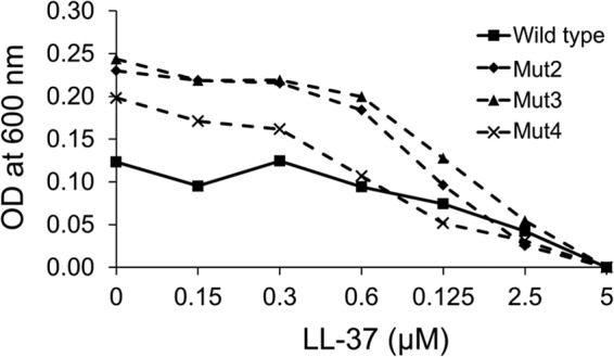
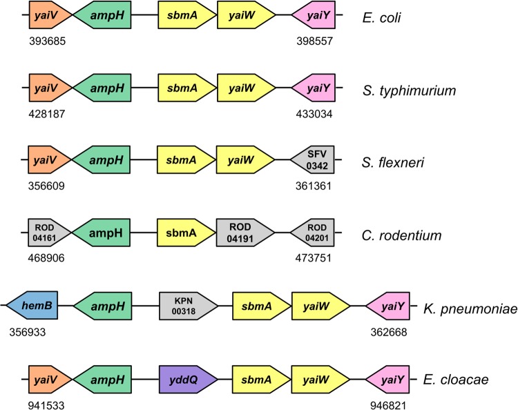
Due to their lack of toxicity to mammalian cells and good serum stability, proline-rich antimicrobial peptides (PR-AMPs) have been proposed as promising candidates for the treatment of infections caused by antimicrobial-resistant bacterial pathogens. It has been hypothesized that these peptides act on multiple targets within bacterial cells, and therefore the likelihood of the emergence of resistance was considered to be low. Here, we show that spontaneous Escherichia coli mutants resistant to pyrrhocoricin arise at a frequency of approximately 6 × 10(-7). Multiple independently derived mutants all contained a deletion in a nonessential gene that encodes the putative peptide uptake permease SbmA. Sensitivity could be restored to the mutants by complementation with an intact copy of the sbmA gene. These findings question the viability of the development of insect PR-AMPs as antimicrobials.


Figures

FIG 1
FIG 1
(A) Sensitivity of wild-type E. coli C600 and Mut1 to Mut4 to pyrrhocoricin. The MIC value was 5 μM for the wild-type bacteria, higher than 80 μM for Mut1, and higher than 640 μM for Mut2, Mut3, and Mut4. (B) Growth curve for wild-type E. coli C600 and highly resistant mutants Mut2 to Mut4.
FIG 2
FIG 2
Analysis of the deletion of the region around the sbmA gene in pyrrhocoricin-resistant E. coli C600 mutants. (A) Genomic structure of the deleted region in Mut2. Nucleotide positions are indicated with reference to the published E. coli K-12 W3110 genome. Gray horizontal arrows show the positions of the forward and reverse PCR primers (Table 1). (B) The deletion boundaries in Mut1, Mut3, and Mut4 are close to but different from those in Mut2. Eight-nucleotide direct repeats that likely mediate the site-specific recombination leading to deletion are shown on top. (C, D) Confirmation of deletion of the sbmA-containing region in Mut1 to Mut4 by PCR. (C) The products of PCR amplification of the sbmA gene from the wild type (lane 1) and mutant E. coli C600 strains (lanes 2 to 5, Mut1, Mut2, Mut3, and Mut4, respectively), using flanking primers sbmA-del-F and sbmA-del-R (Table 1). Expected size of the PCR product was 1.4 kb. (D) PCR-amplified DB-F/DB-R fragments (Table 1). Lane 1 (parental strain), anticipated size (7,096 bp) is consistent with the mobility on the gel; lanes 2 to 5 (Mut1 to Mut4, respectively), size estimation from the gel, 2.4 kb for Mut1, Mut3, and Mut4 and 1.6 kb for Mut2. The PCR products for all mutants were sequenced and were shown to be 2,368 bp for Mut1, Mut3, and Mut4 (due to a 4,728-bp deletion) and 1,631 bp for Mut2 (due to a 5,465-bp deletion).
FIG 3
FIG 3
Confirmation of expression of SbmA (46.5 kDa) from a multicopy plasmid pET-22b(+) in the complemented deletion mutant Mut2. Fifteen micrograms of total protein from the uninduced (−IPTG) or induced (+IPTG) samples was loaded per lane.
FIG 4
FIG 4
Sensitivity to pyrrhocoricin of Mut2 to Mut4 genetically complemented with an IPTG-inducible high-copy-number plasmid expressing SbmA (solid line). Sensitivity of noncomplemented mutants is shown in a dashed line for comparison. Expression of the transporter largely restored sensitivity to pyrrhocoricin.
FIG 5
FIG 5
Mutations in the sbmA gene sustained by the recombinant sbmA-pET-22b(+) plasmid when gene expression was induced in the presence of pyrrhocoricin. (A) Disruption of the sbmA coding region by the 768-bp transposon insertion (E. coli insertion sequence 1 [IS1]) between positions 956 and 964 in clones I-Mut2-1 and IMut2-2. The sequences of 9-bp direct repeats of the target sequence, originating from the insertion, and the 23-bp inverted repeats at the IS1 termini are shown. (B) sbmA gene disruption by a sequence of unknown origin inserted in the coding sequence before nucleotide 770 in clone I-Mut3-1. (C) The IS1 transposon inserted between positions 1004 and 1012 of the sbmA coding region in clones II-Mut3-2 and II-Mut3-3. (D) A single-base deletion in sbmA identified in clone III-Mut3-4.
FIG 6
FIG 6
Sensitivity to LL-37 of wild-type E. coli C600 and Mut2 to Mut4. The MIC value was 5 μM for both the wild-type bacteria and mutants.
FIG 7
FIG 7
Schematic representation of the genomic regions adjacent to sbmA in E. coli K-12 W3110 (NCBI NC_007779.1) (26), Salmonella enterica serovar Typhimurium strain LT2 (NCBI NC_003197.1) (52), Shigella flexneri 5 8401 (NCBI NC_008258.1) (53), Citrobacter rodentium ICC168 (GenBank FN543502.1) (54), Klebsiella pneumoniae MGH 78578 (Genome Sequencing Center of Washington University, http://genome.wustl.edu/genomes/view/klebsiella_pneumoniae/; GenBank accession no. CP000647.1), Enterobacter cloacae EcWSU1 (GenBank accession no. CP002886.1) (55). Numbers show the location in the genome. Arrows indicate gene transcription direction. One color for sbmA and yaiW (yellow) indicates that they form an operon.

References

    1. Centers for Disease Control and Prevention (CDC). 2013. Antibiotic resistance threats in the United States, 2013. CDC, Atlanta, GA: http://www.cdc.gov/drugresistance/threat-report-2013
    1. Hancock REW. 2007. The end of an era. Nat. Rev. Drug Discov. 6:28. 10.1038/nrd2223 - DOI
    1. Ho J, Tambyah PA, Paterson DL. 2010. Multiresistant Gram-negative infections: a global perspective. Curr. Opin. Infect. Dis. 23:546–553. 10.1097/QCO.0b013e32833f0d3e - DOI - PubMed
    1. Scocchi M, Tossi A, Gennaro R. 2011. Proline-rich antimicrobial peptides: converging to a non-lytic mechanism of action. Cell. Mol. Life Sci. 68:2317–2330. 10.1007/s00018-011-0721-7 - DOI - PMC - PubMed
    1. Otvos L., Jr 2002. The short proline-rich antibacterial peptide family. Cell. Mol. Life Sci. 59:1138–1150. 10.1007/s00018-002-8493-8 - DOI - PMC - PubMed

Publication types

MeSH terms

Substances

LinkOut - more resources