

Alpha-synuclein mRNA expression in oligodendrocytes in MSA
- PMID: 24590631
- PMCID: PMC4238782
- DOI: 10.1002/glia.22653
Alpha-synuclein mRNA expression in oligodendrocytes in MSA
Abstract
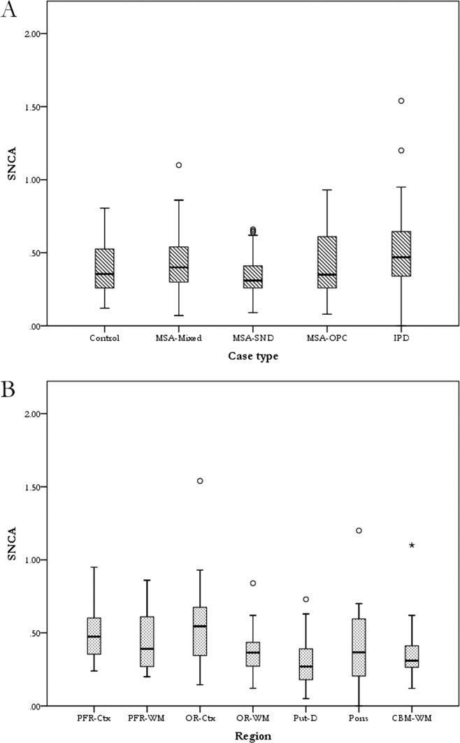
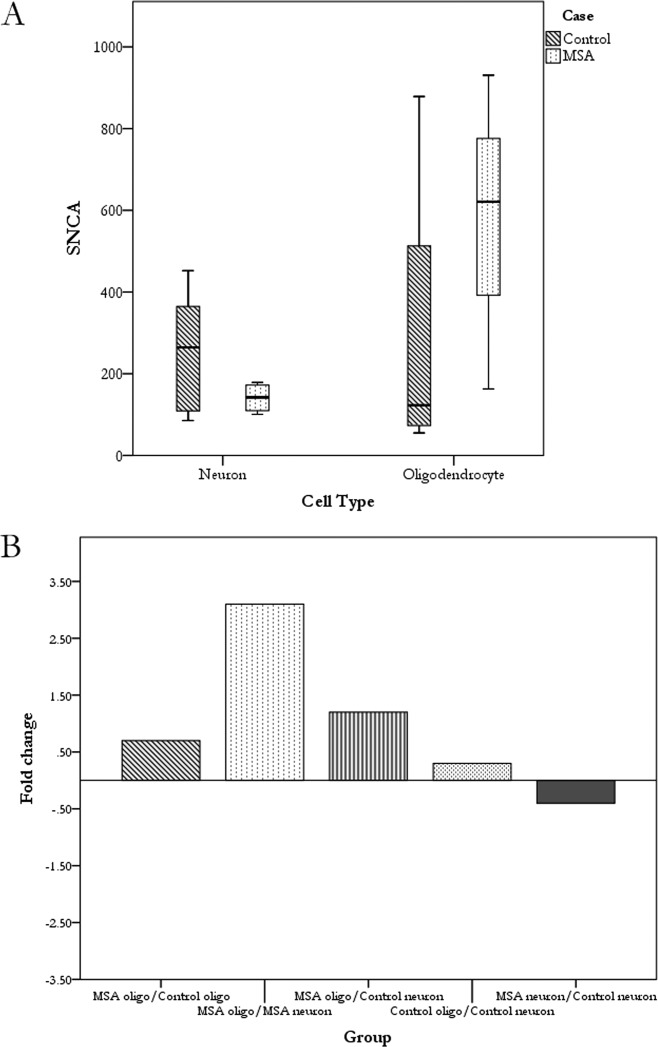
Multiple system atrophy (MSA) is a progressive neurodegenerative disease presenting clinically with parkinsonian, cerebellar, and autonomic features. α-Synuclein (αsyn), encoded by the gene SNCA, is the main constituent of glial cytoplasmic inclusion (GCI) found in oligodendrocytes in MSA, but the methods of its accumulation have not been established. The aim of this study is to investigate alterations in regional and cellular SNCA mRNA expression in MSA as a possible substrate for GCI formation. Quantitative reverse transcription polymerase chain reaction (qPCR) was performed on postmortem brain samples from 15 MSA, 5 IPD, and 5 control cases to investigate regional expression in the frontal and occipital regions, dorsal putamen, pontine base, and cerebellum. For cellular expression analysis, neurons and oligodendrocytes were isolated by laser-capture microdissection from five MSA and five control cases. SNCA mRNA expression was not significantly different between the MSA, IPD and control cases in all regions (multilevel model, P = 0.14). After adjusting for group effect, the highest expression was found in the occipital cortex while the lowest was in the putamen (multilevel model, P < 0.0001). At the cellular level, MSA oligodendrocytes expressed more SNCA than control oligodendrocytes and expression in MSA neurons was slightly lower than that in controls, however, these results did not reach statistical significance. We have demonstrated regional variations in SNCA expression, which is higher in cortical than subcortical regions. This study is the first to demonstrate SNCA mRNA expression by oligodendrocytes in human postmortem tissue using qPCR and, although not statistically significant, could suggest that this may be increased in MSA compared to controls.
Keywords: glial cytoplasmic inclusions; laser-capture microdissection; multiple system atrophy; oligodendrocytes; α-synuclein.
© 2014 The Authors. Glia Published by Wiley Periodicals, Inc.
Figures
References
-
- Ahmed Z, Asi YT, Sailer A, Lees AJ, Houlden H, Revesz T, Holton JL. The neuropathology, pathophysiology and genetics of multiple system atrophy. Neuropathol Appl Neurobiol. 2012;38:4–24. - PubMed
-
- Beyer K, Domingo-Sàbat M, Humbert J, Carrato C, Ferrer I, Ariza A. Differential expression of alpha-synuclein, parkin, and synphilin-1 isoforms in Lewy body disease. Neurogenetics. 2008;9:163–172. - PubMed
-
- Culvenor JG, Rietze RL, Bartlett PF, Masters CL, Li Q-X. Oligodendrocytes from neural stem cells express alpha-synuclein: increased numbers from presenilin 1 deficient mice. Neuroreport. 2002;13:1305–1308. - PubMed
Publication types
MeSH terms
Substances
Grants and funding
LinkOut - more resources
Full Text Sources
Other Literature Sources
Miscellaneous
