. 2014 May;23(5):639-51.
doi: 10.1002/pro.2448. Epub 2014 Mar 26.

Patterns of structural dynamics in RACK1 protein retained throughout evolution: a hydrogen-deuterium exchange study of three orthologs

Affiliations

Patterns of structural dynamics in RACK1 protein retained throughout evolution: a hydrogen-deuterium exchange study of three orthologs

Krzysztof Tarnowski et al. Protein Sci. 2014 May.

Abstract

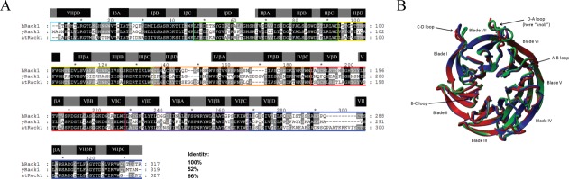
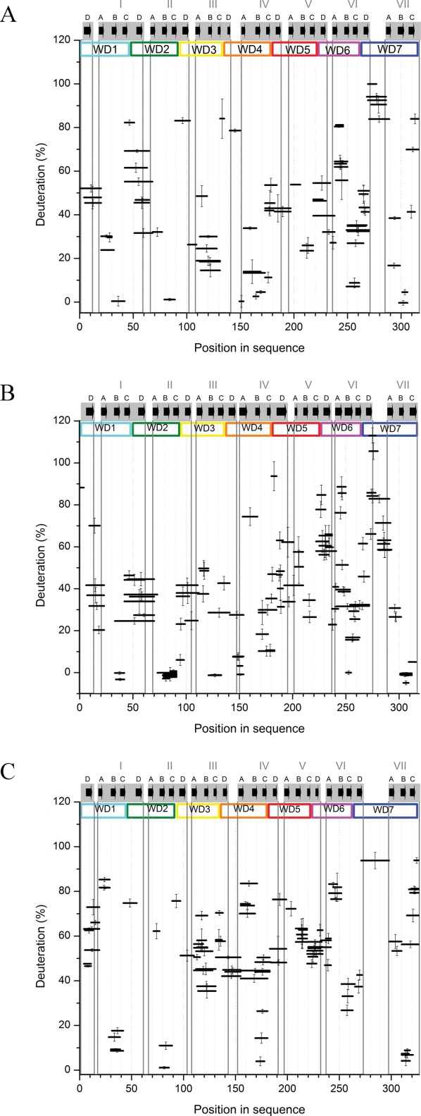
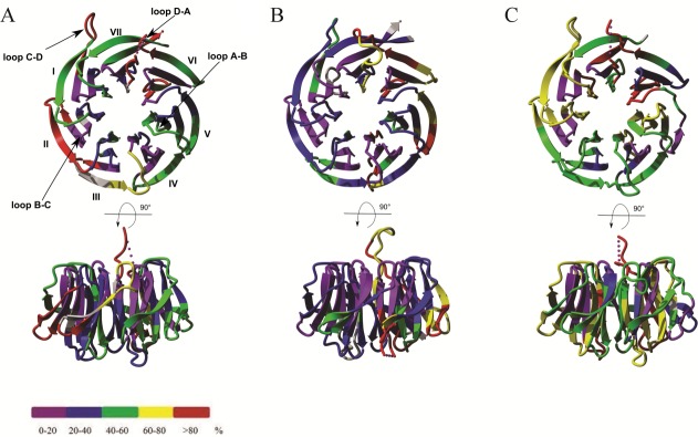
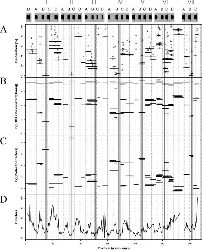
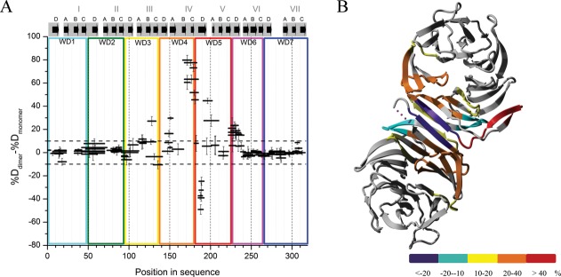
RACK1 is a member of the WD repeat family of proteins and is involved in multiple fundamental cellular processes. An intriguing feature of RACK1 is its ability to interact with at least 80 different protein partners. Thus, the structural features enabling such interactomic flexibility are of great interest. Several previous studies of the crystal structures of RACK1 orthologs described its detailed architecture and confirmed predictions that RACK1 adopts a seven-bladed β-propeller fold. However, this did not explain its ability to bind to multiple partners. We performed hydrogen-deuterium (H-D) exchange mass spectrometry on three orthologs of RACK1 (human, yeast, and plant) to obtain insights into the dynamic properties of RACK1 in solution. All three variants retained similar patterns of deuterium uptake, with some pronounced differences that can be attributed to RACK1's divergent biological functions. In all cases, the most rigid structural elements were confined to B-C turns and, to some extent, strands B and C, while the remaining regions retained much flexibility. We also compared the average rate constants for H-D exchange in different regions of RACK1 and found that amide protons in some regions exchanged at least 1000-fold faster than in others. We conclude that its evolutionarily retained structural architecture might have allowed RACK1 to accommodate multiple molecular partners. This was exemplified by our additional analysis of yeast RACK1 dimer, which showed stabilization, as well as destabilization, of several interface regions upon dimer formation.

Keywords: WD repeats; cell signaling; hydrogen deuterium exchange; mass spectrometry; protein dynamics; receptor for activated C kinase; scaffolding protein.


Figures

Figure 1
Figure 1
RACK1 orthologs. (A) Sequence alignment of RACK1 orthologs from H. sapiens (hRACK1), S. cerevisiae (yRACK1), and A. thaliana, isoform A (atRACK1). Percentage sequence identity is shown at end of the alignment. Characteristic WD repeats are highlighted with colored rectangles. β-propeller blades are highlighted by gray rectangles that contain A, B, C, D β-strands (black rectangles), as assigned based on hRACK1crystal structures available in PDB (4AOW). (B) Superposition of the crystal structures of hRACK1 (4AOW, blue), yRACK1 (3FRX, green,) and atRACK1 (3DM0, red). Characteristic structural elements such as blades and loops are highlighted.
Figure 2
Figure 2
Percentage of deuteration of peptic fragments from human (A), yeast (B), and plant (C) RACK1 at 10 s exchange time. Position of a peptide in the sequence is shown on the horizontal axis, represented by a horizontal bar with length equal to the length of the peptide. Position of the bar at the vertical axis marks the fraction exchanged after 10 s. y-Axis error bars are standard deviations calculated from three independent experiments. WD repeats are marked and colored as in Figure 1(A). β-Propeller blades are highlighted by gray rectangles that contain A, B, C, and D β-strands (black arrows), as assigned based on RACK1 crystal structures available in PDB (4AOW, 3RFH, and 3DM0).
Figure 3
Figure 3
Overlay of 10 s hydrogen-deuterium exchange results on crystal structures from (A) hRACK1 (4AOW), (B) yRACK1 (3FRX), and (C) atRACK1 (3DM0) at 10 s exchange time. Structures are color coded according to HDXMS results of the shortest available peptide: violet, strongly protected (0–20%); blue, protected (20–40%); green, moderately protected (40–60%); yellow, moderately flexible (60–80%); and red, flexible (>80%). Gray color represents regions not covered by peptic peptides in the sequence.
Figure 4
Figure 4
Regions of fast and slow hydrogen-deuterium exchange in hRACK1, correlated with B-factor of its crystal structure. The gray shaded regions represent B-C loops alone. (A) Percentage of deuteration of peptic fragments from hRACK1 at 10 s [horizontal bars as in Fig. 2(A)], 1 min (open triangles), 20 min (filled circles), and 1 h (open circles) of exchange. (B) average rate of exchange kex (log scale) obtained by a monoexponential fit for each peptide, marked by the position of a black horizontal bar with length equal to the length of the peptide on the vertical axis. Position of each peptide in the sequence is shown on the horizontal axis. Intrinsic rates of exchange kint for each peptide are also shown marked by gray bars. Note a few peptides, characterized by low kex values, leading to very high protection factor (P) values, as shown in log scale in (C). (D) B-factor value profile for hRACK1 crystal structure, with several minima correlating with regions of lowest kex and highest P values.
Figure 5
Figure 5
Differences in fraction of exchanged amide protons for peptic peptides between yRACK1 monomers and dimers. (A) Subtraction plot representing differences in exchange between peptides of dimeric and monomeric yRACK1. WD repeats, blades, and strands of β-propeller are marked as in Figure 2(B). (B) Differences in amide proton exchange between monomeric and dimeric yRACK1 overlaid onto crystal structure of its dimer (3RFH). Differences upon dimer formation are color coded as follows: violet, strong stabilization (less than −20%); cyan, stabilization (−20 to −10%); yellow, weak destabilization (10 to 20%); orange, moderate destabilization (20 to 40%); and red, high destabilization (more than 40%).

References

    1. Mochly-Rosen D, Khaner H, Lopez J. Identification of intracellular receptor proteins for activated protein kinase C. Proc Natl Acad Sci USA. 1991;88:3997–4000. - PMC - PubMed
    1. Schloss JA. A Chlamydomonas gene encodes a G protein beta subunit-like polypeptide. Mol Gen Genet. 1990;221:443–452. - PubMed
    1. Kuo WN, Jones DL, Ku TW, Weeks KD, Jordon PM, Dopson NC. Immunoreactivity of PKC gammalambda and RACK1 in baker's yeast, lobster and wheat germ. Biochem Mol Biol Int. 1995;36:957–963. - PubMed
    1. Kwak JM, Kim SA, Lee SK, Oh SA, Byoun CH, Han JK, Nam HG. Insulin-induced maturation of Xenopus oocytes is inhibited by microinjection of a Brassica napus cDNA clone with high similarity to a mammalian receptor for activated protein kinase C. Planta. 1997;201:245–251. - PubMed
    1. Vani K, Yang G, Mohler J. Isolation and cloning of a Drosophila homolog to the mammalian RACK1 gene, implicated in PKC-mediated signalling. Biochim Biophys Acta. 1997;1358:67–71. - PubMed

Publication types

MeSH terms