. 2014 Apr 18;289(16):11421-11430.
doi: 10.1074/jbc.M113.537498. Epub 2014 Mar 3.

Adenovirus composition, proteolysis, and disassembly studied by in-depth qualitative and quantitative proteomics

Affiliations

Adenovirus composition, proteolysis, and disassembly studied by in-depth qualitative and quantitative proteomics

Marco Benevento et al. J Biol Chem. .

Abstract

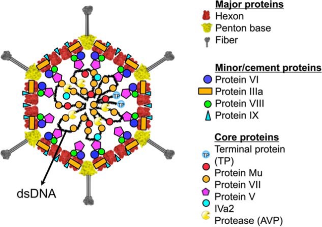
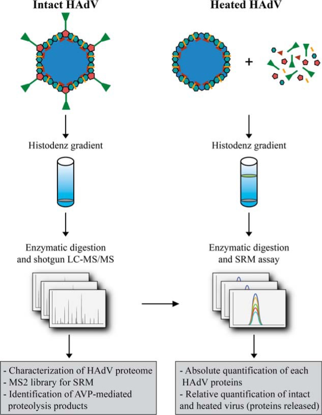
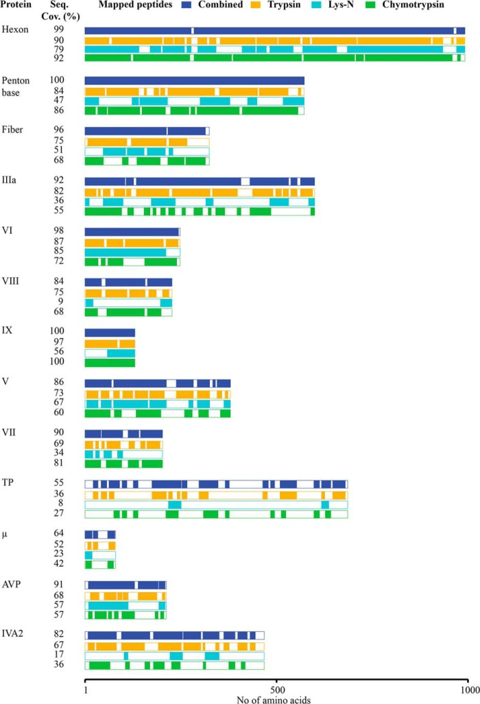
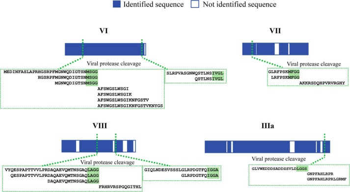
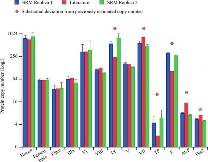
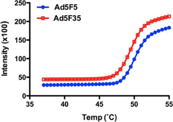
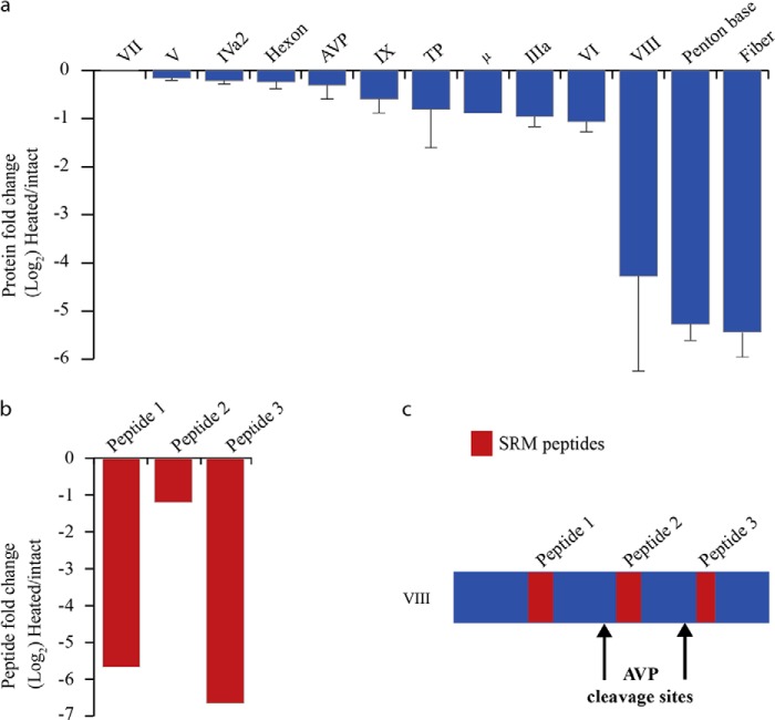
Using high-resolution MS-based proteomics in combination with multiple protease digestion, we profiled, with on average 90% sequence coverage, all 13 viral proteins present in an human adenovirus (HAdV) vector. This in-depth profile provided multiple peptide-based evidence on intrinsic protease activity affecting several HAdV proteins. Next, the generated peptide library was used to develop a targeted proteomics method using selected reaction monitoring (SRM) aimed at quantitative profiling of the stoichiometry of all 13 proteins present in the HAdV. We also used this method to probe the release of specific virus proteins initiated by thermal stimulation, mimicking the early stage of HAdV disassembly during entry into host cells. We confirmed the copy numbers of the most well characterized viral capsid components and established the copy numbers for proteins whose stoichiometry has so far not been accurately defined. We also found that heating HAdV induces the complete release of the penton base and fiber proteins as well as a substantial release of protein VIII and VI. For these latter proteins, maturational proteolysis by the adenoviral protease leads to the differential release of fragments with certain peptides being fully released and others largely retained in the AdV particles. This information is likely to be beneficial for the ongoing interpretation of high resolution cryoEM and x-ray electron density maps.

Keywords: Adenovirus; Mass Spectrometry (MS); Proteomics; Virus Entry; Virus Structure.


Figures

FIGURE 1.
FIGURE 1.
Structural model of adenovirus (51). Schematic diagram of HAdV showing the structural proteins for which the localization in the virion is known. The major capsid proteins are the hexon, penton base, and fiber. The capsid is stabilized on the outside by the minor protein IX and IIIa (A. Reddy and P. Nemerow, personal communication). The remaining minor capsid proteins, VI and VIII, have been localized to the inner surface of the capsid. The core proteins, TP, Mu, V, and VII, are associated with the double-stranded DNA genome in the interior of the virion. The presence and locations of AVP and the protein IVa2 are currently ambiguous.
FIGURE 2.
FIGURE 2.
Schematic of the MS-based workflow combining shotgun and targeted approaches to characterize HAdV (Ad5F35). HAdV from intact and heat-stressed particles were purified by HistoDenz density gradients. A first analysis was performed by combining shotgun LC-MS/MS analysis with multiple proteases for protein digestion. This analysis was used to identify all HAdV proteins with high sequence coverage, identify AVP cleavage sites, and build a spectral library from which we selected the peptides employed in the SRM assay. Using these assays, we quantitatively profiled the stoichiometry of all 13 proteins present, as well as their (partial) release upon heat stress.
FIGURE 3.
FIGURE 3.
High-resolution MS-based proteomics profiling in combination with multiple protease digestion allowed sequence coverage of on average 90% for the HAdV proteome. Representation of the 13 HAdV proteins identified in the shotgun LC-MS/MS analyses. The sequence coverage (%) along with the specific area of the protein sequenced is reported for each of the employed enzymes trypsin (orange), Lys-N (light blue), and chymotrypsin (green), as well as the cumulative coverage (dark blue).
FIGURE 4.
FIGURE 4.
Identified AVP cleavage sites in four HAdV (Ad5F35) proteins. Representation of the four HAdV proteins (bars) for which direct evidence of AVP cleavage could be found. For each of the six cleavage sites, the set of confidently identified peptides deriving from multiple protease digestions are highlighted in the green boxes.
FIGURE 5.
FIGURE 5.
Stoichiometry of the proteins in HAdV (Ad5F35) particles. The bar chart reports the copy number (log2) for each of the HAdV proteins obtained by the SRM assay in two independent biological replicates (blue and red bars) and the estimated copy number known from literature (green bars). The error bars indicate the relative S.D. calculated from the quantitative values, retrieved by each of the SRM peptides employed for each protein.
FIGURE 6.
FIGURE 6.
Thermally induced disassembly of AdV capsids. Ad5F5 (HAdV5) and Ad5F35 were heated to the indicated temperatures in the presence of Pico green. Capsid disassembly, as measured by dye accessibility to the viral genome, was measured in relative fluorescence units. Data are the mean ± S.D. from triplicate samples. From the data, it appears that Ad5F5 and Ad5F35 disassemble with similar kinetics, and both reach a plateau at ∼55 °C.
FIGURE 7.
FIGURE 7.
Quantification of the released HAdV proteins upon heat stress. a, the fold change in the abundance of HAdV proteins upon heat stress are shown. The y axis represents the protein ratios of heat-stressed/intact virus in log2 scale, and the error bars represent the relative S.D. b, fold change in the abundance of various segments of protein VIII, generated by the HAdV protease: peptides 1 and 3 are fully released, whereas peptide 2 is only marginally released. c, the protein domain in which these peptides reside suggest that only the N- and C-terminal segment of protein VIII are completely released.

References

    1. Davison A. J., Benko M., Harrach B. (2003) Genetic content and evolution of adenoviruses. J. Gen. Virol. 84, 2895–2908 - PubMed
    1. Reddy V. S., Natchiar S. K., Stewart P. L., Nemerow G. R. (2010) Crystal structure of human adenovirus at 3.5 Å resolution. Science 329, 1071–1075 - PMC - PubMed
    1. Liu H., Jin L., Koh S. B., Atanasov I., Schein S., Wu L., Zhou Z. H. (2010) Atomic structure of human adenovirus by cryo-EM reveals interactions among protein networks. Science 329, 1038–1043 - PMC - PubMed
    1. Nemerow G. R. (2000) Cell receptors involved in adenovirus entry. Virology 274, 1–4 - PubMed
    1. Chow L. T., Gelinas R. E., Broker T. R., Roberts R. J. (1977) An amazing sequence arrangement at the 5′ ends of adenovirus 2 messenger RNA. Cell 12, 1–8 - PubMed

Publication types