Review
. 2014 Mar 1;4(3):a014076.
doi: 10.1101/cshperspect.a014076.

Myocardial tissue engineering: in vitro models

Affiliations
Review

Myocardial tissue engineering: in vitro models

Gordana Vunjak Novakovic et al. Cold Spring Harb Perspect Med. .

Abstract

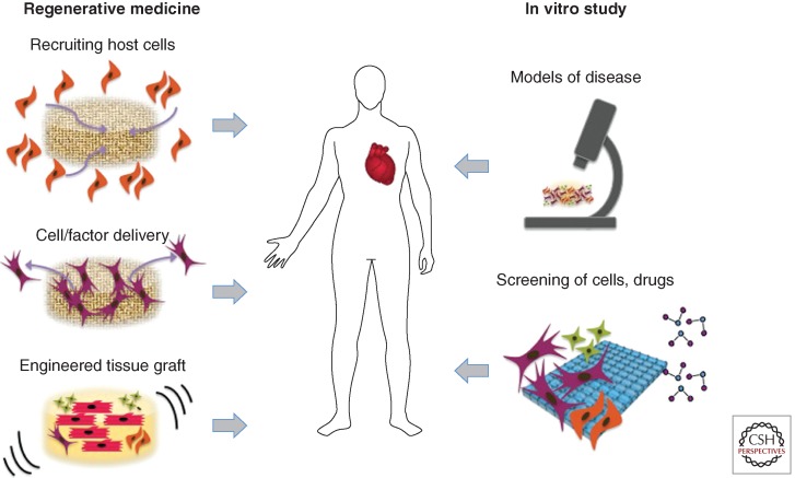
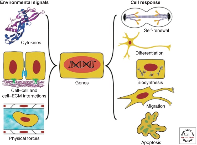
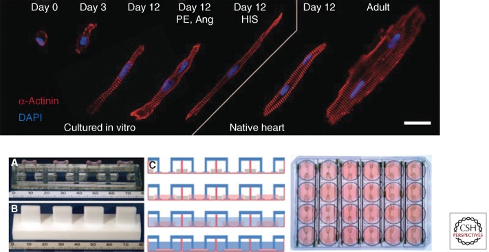
Modeling integrated human physiology in vitro is a formidable task not yet achieved with any of the existing cell/tissue systems. However, tissue engineering is becoming increasingly successful at authentic representation of the actual environmental milieu of tissue development, regeneration and disease progression, and in providing real-time insights into morphogenic events. Functional human tissue units engineered to combine biological fidelity with the high-throughput screening and real-time measurement of physiological responses are poised to transform drug screening and predictive modeling of disease. In this review, we focus on the in vitro engineering of functional human myocardium that mimics heart tissue for analysis of myocardial function, in the context of physiological studies, drug screening for therapeutics, and safety pharmacology.


Figures

Figure 1.
Figure 1.
Myocardial tissue engineering. The classical paradigm of myocardial tissue engineering involves cultivation of cells on scaffolds, with the application of molecular and physical factors (via bioreactors), for implantation. Alternatively, host cells can be recruited to the repair site by implanted scaffolds, with or without cells. More recently, myocardial tissue engineering is being increasingly used for modeling disease and in high-throughput platforms for screening of drugs and therapeutic targets.
Figure 2.
Figure 2.
Environmental regulation of cell function. Both in vivo and in vitro, cells interact with the entire context of their environment: cytokines, surrounding cells, extracellular matrix, and physical forces (hydrodynamic, mechanical, electrical). In response to the combined effects of these factors, the cells may take a number of different fates—from differentiation to apoptosis and death. At the same time, the cells modulate their environment by secreting cytokines, remodeling the extracellular matrix, affecting the surrounding cells, and generating forces. Cardiomyocytes are an example of the cells that actively interact with their environment throughout development, adult life, as well as under pathological conditions. The designs of advanced systems for myocardial tissue engineering are “biomimetic” in nature as they recapitulate key regulatory factors, molecular and physical, acting in vivo. (From Vunjak-Novakovic and Scadden 2011; reproduced, with permission, from the author.)
Figure 3.
Figure 3.
In vitro testing platforms. (Top) Engineered cardiac tissues can reach the level of advanced morphologic differentiation and maturation typical of their age-matched in vivo counterparts. Hypertrophic cardiomyocyte growth in hydrogel with the application of mechanical stretch is compared to native heart. Cardiomyocytes from engineered tissues at culture days 0, 3, and 12 from three experimental groups: (1) untreated, (2) treated with phenylephrine (PE, 20 µmol/L), angiotensin II (Ang, 100 nmol/L), and (3) treated with hypertrophy-inducing serum (HIS) are compared to cardiomyocytes from rat myocardium. Red, α-actinin; blue, DAPI-labeled nuclei. Scale bar, 20 µm. The image is an assembly of individual immunostains of representative cells from each group. (From Tiburcy et al. 2011; reproduced, with permission, from the author.) (Bottom) Experimental setup for casting and cultivation of cardiac tissue. (A) Silicone post rack with four tissues, turned upside down, scale in mm. (B) Teflon spacer to generate agarose molds, turned upside down, scale in mm. (C) Generation of cardiac tissues. First lane, Casting molds are generated in 24-well plates using agarose and Teflon spacers. Silicone racks are placed on the dish, pairs of posts reach into each mold. Second lane, Cell suspension in Matrigel, fibrinogen, and thrombin is pipetted into the molds. Third lane, Two hours later, the hydrogel is polymerized, with silicone posts embedded in hydrogel at both ends. The constructs are removed from the molds and transferred to 24-well plates. Fourth lane, The constructs are maintained in culture for 15 to 30 days. (From Hansen et al. 2010; reproduced, with permission, from the author.)

References

    1. Atala A, Bauer SB, Soker S, Yoo JJ, Retik AB 2006. Tissue-engineered autologous bladders for patients needing cystoplasty. The Lancet 367: 1241–1246 - PubMed
    1. Birla RK, Borschel GH, Dennis RG, Brown DL 2005. Myocardial engineering in vivo: Formation and characterization of contractile, vascularized three-dimensional cardiac tissue. Tissue Eng 11: 803–813 - PubMed
    1. Boudou T, Legant WR, Mu A, Borochin MA, Thavandiran N, Radisic M, Zandstra PW, Epstein JA, Margulies KB, Chen CS 2012. A microfabricated platform to measure and manipulate the mechanics of engineered cardiac microtissues. Tissue Eng Part A 18: 910–919 - PMC - PubMed
    1. Buckingham M, Meilhac S, Zaffran S 2005. Building the mammalian heart from two sources of myocardial cells. Nat Rev Genet 6: 826–835 - PubMed
    1. Bursac N, Papadaki M, Cohen RJ, Schoen FJ, Eisenberg SR, Carrier R, Vunjak-Novakovic G, Freed LE 1999. Cardiac muscle tissue engineering: Towards an in vitro model for electrophysiological studies. Am J Physiol 277: H433–H444 - PubMed

Substances

LinkOut - more resources