. 2014 Mar 18;111(11):E1035-42.
doi: 10.1073/pnas.1401595111. Epub 2014 Mar 3.

Blood-spinal cord barrier disruption contributes to early motor-neuron degeneration in ALS-model mice

Affiliations

Blood-spinal cord barrier disruption contributes to early motor-neuron degeneration in ALS-model mice

Ethan A Winkler et al. Proc Natl Acad Sci U S A. .

Abstract

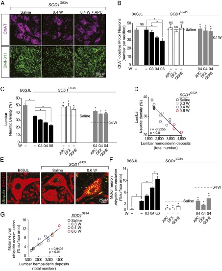
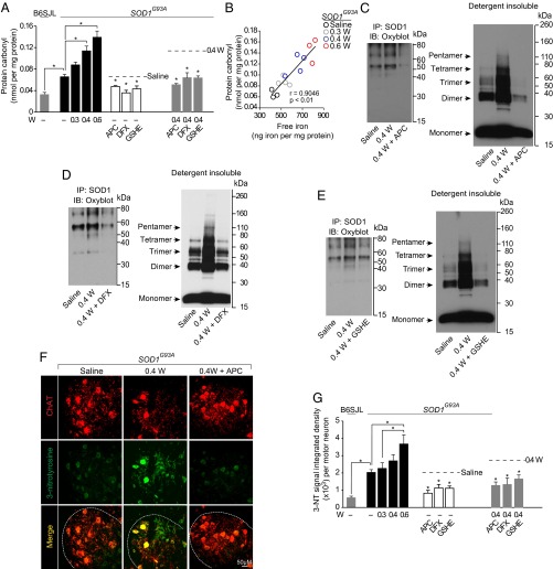
Humans with ALS and transgenic rodents expressing ALS-associated superoxide dismutase (SOD1) mutations develop spontaneous blood-spinal cord barrier (BSCB) breakdown, causing microvascular spinal-cord lesions. The role of BSCB breakdown in ALS disease pathogenesis in humans and mice remains, however, unclear, although chronic blood-brain barrier opening has been shown to facilitate accumulation of toxic blood-derived products in the central nervous system, resulting in secondary neurodegenerative changes. By repairing the BSCB and/or removing the BSCB-derived injurious stimuli, we now identify that accumulation of blood-derived neurotoxic hemoglobin and iron in the spinal cord leads to early motor-neuron degeneration in SOD1(G93A) mice at least in part through iron-dependent oxidant stress. Using spontaneous or warfarin-accelerated microvascular lesions, motor-neuron dysfunction and injury were found to be proportional to the degree of BSCB disruption at early disease stages in SOD1(G93A) mice. Early treatment with an activated protein C analog restored BSCB integrity that developed from spontaneous or warfarin-accelerated microvascular lesions in SOD1(G93A) mice and eliminated neurotoxic hemoglobin and iron deposits. Restoration of BSCB integrity delayed onset of motor-neuron impairment and degeneration. Early chelation of blood-derived iron and antioxidant treatment mitigated early motor-neuronal injury. Our data suggest that BSCB breakdown contributes to early motor-neuron degeneration in ALS mice and that restoring BSCB integrity during an early disease phase retards the disease process.

Keywords: amyotrophic lateral sclerosis; neurodegeneration.


Conflict of interest statement

Conflict of interest statement: B.V.Z. is the scientific founder of ZZ Biotech, a start-up biotechnology company that is developing an activated protein C analog for stroke with possible implications in other neurological disorders. J.H.G. is a member of the Scientific Advisory Board of ZZ Biotech.

Figures

Fig. 1.
Fig. 1.
Early microvascular lesions in SOD1G93A mice: acceleration by Warfarin and protection by 5A-APC. (A–E) IgG (A and B), hemoglobin (C), hemosiderin (D), and free iron (E) in the lumbar anterior horn of SOD1G93A mice receiving from 35 to 95 d saline, 0.3–0.6 mg⋅kg−1⋅d−1 warfarin (W), and/or 100 µg⋅kg−1⋅d−1 5A-APC (APC), 100 mg⋅kg−1⋅d−1 deferoxamine mesylate (DFX), or 50 mg⋅kg−1⋅d−1 glutathione monoethyl ester (GSHE) with either saline or warfarin (0.4 mg⋅kg−1⋅d−1). B6SJL littermates and SOD1WT mice received saline or warfarin (0.6 mg⋅kg−1⋅d−1). (F, Upper) Prussian blue-positive hemosiderin deposits (blue) and collagen IV-positive capillaries (brown) in lumbar cord anterior horn of 95-d-old B6SJL littermate and saline-treated SOD1G93A mouse. (Lower) Perl’s-positive hemosiderin deposits (red) in 95-d-old SOD1G93A mouse receiving saline or 0.6 mg⋅kg−1⋅d−1 warfarin. Broken line denotes boundary of the anterior horn. (G) Collagen IV-positive capillaries (green) and CD235a-positive erythrocytes (red) in the lumbar anterior horn of 95-d-old B6SJL littermate receiving 0.6 mg⋅kg−1⋅d−1 warfarin and SOD1G93A mice receiving saline, 0.4 mg⋅kg−1⋅d−1 warfarin or 0.4 mg⋅kg−1⋅d−1 warfarin and 5A-APC (100 μg⋅kg−1⋅d−1). (H) Percentage of capillary-associated and noncapillary hemosiderin deposits in 95-d-old SOD1G93A mice receiving saline or warfarin (0.6 mg⋅kg−1⋅d−1); n = 4–5 mice per group. (I) Positive correlation between the number of lumbar hemosiderin deposits and the degree of anticoagulation determined as international normalized ratio (INR) in 95-d-old SOD1G93A mice receiving saline or warfarin (0.3–0.6 mg⋅kg−1⋅d−1). Individual data points from four to five animals per group; r, Pearson’s correlation. In B–E, mean ± SEM, n = 3–5 mice per group. *P < 0.05; NS, nonsignificant. In B–E, APC, DFX, and GSHE treatments were compared with the respective saline or warfarin treatments as indicated by broken lines.
Fig. 2.
Fig. 2.
Onset of motor impairment and prevention in SOD1G93A mice with spontaneous and accelerated microvascular lesions. (A and B) Cumulative probability (A) and mean age (B) of motor impairment in SOD1G93A mice treated with saline (n = 14) or 0.3 (n = 21), 0.4 (n = 15), and 0.6 (n = 16) mg⋅kg−1⋅d−1 warfarin (W) from day 35 postnatal. Acceleration of motor symptoms in days relative to saline is provided above each group. Values in B, mean ± SEM, *P < 0.05. Nontransgenic B6SJL littermates (n = 14) received 0.6 mg⋅kg−1⋅d−1 warfarin. (C) Negative correlation between onset of motor impairment and number of lumbar hemosiderin deposits in SOD1G93A mice treated with saline or warfarin (0.3–0.6 mg⋅kg−1⋅d−1). Individual data points are from four to five mice per group; r, Pearson’s correlation. (D and E) Cumulative probability (D) and mean age (E) of motor impairment in SOD1G93A mice treated daily with saline (n = 14), 100 µg/kg 5A-APC (n = 15), 100 mg/kg DFX (n = 14), or 50 mg/kg GSHE (n = 14) from day 35 postnatal. Delay of motor symptoms in days relative to saline is provided above each group. Values in E, mean ± SEM, *P < 0.05 compared with saline. Nontransgenic B6SJL littermates (n = 14 per group) received APC, DFX, or GSHE. (F and G) Cumulative probability (F) and mean age (G) of motor impairment in SOD1G93A mice treated with 0.4 mg⋅kg−1⋅d−1 warfarin (n = 15) and 100 µg⋅kg−1⋅d−1 5A-APC (n = 14), 100 mg⋅kg−1⋅d−1 DFX (n = 14), or 50 mg⋅kg−1⋅d−1 GSHE (n = 14) from day 35 postnatal. Delay of motor symptoms in days relative to 0.4 mg/kg warfarin is provided above each group. Values in G, mean ± SEM, *P < 0.05 compared with 0.4 mg⋅kg−1⋅d−1 warfarin.
Fig. 3.
Fig. 3.
Early motor-neuron degenerative changes and prevention in SOD1G93A mice with spontaneous and accelerated microvascular lesions. (A–C) ChAT-positive motor neurons (magenta) and SMI-311–positive neurites (green) (A) and quantification of motor neurons (B) and neuritic density (C) in lumbar spinal cord of 95-d-old nontransgenic littermates and SOD1G93A mice treated with saline, 0.3–0.6 mg⋅kg−1⋅d−1 warfarin (W), or 100 µg⋅kg−1⋅d−1 5A-APC, 100 mg⋅kg−1⋅d−1 DFX, or 50 mg⋅kg−1⋅d−1 GSHE with saline or 0.4 mg⋅kg−1⋅d−1 warfarin. (D) Negative correlation between SMI-311–positive neurites and lumbar hemosiderin deposits of 95-d-old individual SOD1G93A mice treated with saline or 0.3–0.6 mg⋅kg−1⋅d−1 warfarin. r, Pearson’s correlation; n = 4–5 animals per group. (E) Ubiquitin-positive accumulates (green) in motor neurons (red, visualized with SMI-311) in the lumbar anterior horn in 95-d-old SOD1G93A mice treated with saline or 0.6 mg⋅kg−1⋅d−1 warfarin. B6SJL, a nontransgenic littermate control. (F) Quantification of ubiquitin accumulation in motor neurons in mice from B. (G) Positive correlation between motor-neuron ubiquitin accumulation and the number of lumbar hemosiderin deposits in SOD1G93A mice treated with saline or 0.3–0.6 mg⋅kg−1⋅d−1 warfarin. Each point is an individual data point; r, Pearson’s correlation; n = 3–5 mice per group. In B, C, and F, values are mean ± SEM; n = 3–5 mice per group. *P < 0.05; NS, nonsignificant. In B, C, and F, APC, DFX, and GSHE treatments were compared with the respective saline and warfarin treatments as indicated by broken lines.
Fig. 4.
Fig. 4.
Early oxidant stress and prevention in SOD1G93A mice with spontaneous and accelerated microvascular lesions. (A) Oxidized protein carbonyls in lumbar cords of 95-d-old nontransgenic B6SJL controls and SOD1G93A mice treated with saline, 0.3–0.6 mg⋅kg−1⋅d−1 warfarin (W), or 100 µg⋅kg−1⋅d−1 5A-APC (APC), 100 mg⋅kg−1⋅d−1 DFX, or 50 mg⋅kg−1⋅d−1 GSHE with saline or 0.4 mg⋅kg−1⋅d−1 warfarin. (B) Positive correlation between free iron and protein carbonyls in mice from A. Each point is an individual data point; r, Pearson’s correlation; n = 3–4 mice per group. (C–E) Representative immunoblotting of human oxidized SOD1 detergent-insoluble species (Left) and detergent-insoluble SOD1 aggregates (Right) in lumbar cord of 95-d-old SOD1G93A mice treated with saline, 0.4 mg⋅kg−1⋅d−1 warfarin, or 0.4 mg⋅kg−1⋅d−1 warfarin with 100 µg⋅kg−1⋅d−1 5A-APC (C), 100 mg⋅kg−1⋅d−1 DFX (D), or 50 mg⋅kg−1⋅d−1 GSHE (E). (F) ChAT-positive motor neurons (red) and 3-nitrotyrosine (3-NT)-positive signal (green) in lumbar spinal cord of 95-d-old SOD1G93A mice treated with saline, 0.4 mg⋅kg−1⋅d−1 warfarin or 0.4 mg⋅kg−1⋅d−1 warfarin and 100 μg⋅kg−1⋅d−1 5A-APC. (G) Quantification of 3-NT–positive signal in mice from A. In A and G, mean ± SEM; n = 3–5 mice per group. *P < 0.05; NS, nonsignificant.
Fig. 5.
Fig. 5.
A schematic illustrating how early blood–spinal cord barrier (BSCB) breakdown initiates motor-neuron injury and how BSCB-directed treatments blocking different steps in the BSCB pathogenic cascade can prevent early motor-neuron injury.

References

    1. Zlokovic BV. Neurovascular pathways to neurodegeneration in Alzheimer’s disease and other disorders. Nat Rev Neurosci. 2011;12(12):723–738. - PMC - PubMed
    1. Kiernan MC, et al. Amyotrophic lateral sclerosis. Lancet. 2011;377(9769):942–955. - PubMed
    1. Winkler EA, et al. Blood-spinal cord barrier breakdown and pericyte reductions in amyotrophic lateral sclerosis. Acta Neuropathol. 2013;125(1):111–120. - PMC - PubMed
    1. Henkel JS, Beers DR, Wen S, Bowser R, Appel SH. Decreased mRNA expression of tight junction proteins in lumbar spinal cords of patients with ALS. Neurology. 2009;72(18):1614–1616. - PubMed
    1. Miyazaki K, et al. Disruption of neurovascular unit prior to motor neuron degeneration in amyotrophic lateral sclerosis. J Neurosci Res. 2011;89(5):718–728. - PubMed

Publication types

MeSH terms