. 2014 Mar 18;111(11):E1043-52.
doi: 10.1073/pnas.1400889111. Epub 2014 Mar 3.

Fatty acid-inducible ANGPTL4 governs lipid metabolic response to exercise

Affiliations

Fatty acid-inducible ANGPTL4 governs lipid metabolic response to exercise

Milène Catoire et al. Proc Natl Acad Sci U S A. .

Abstract

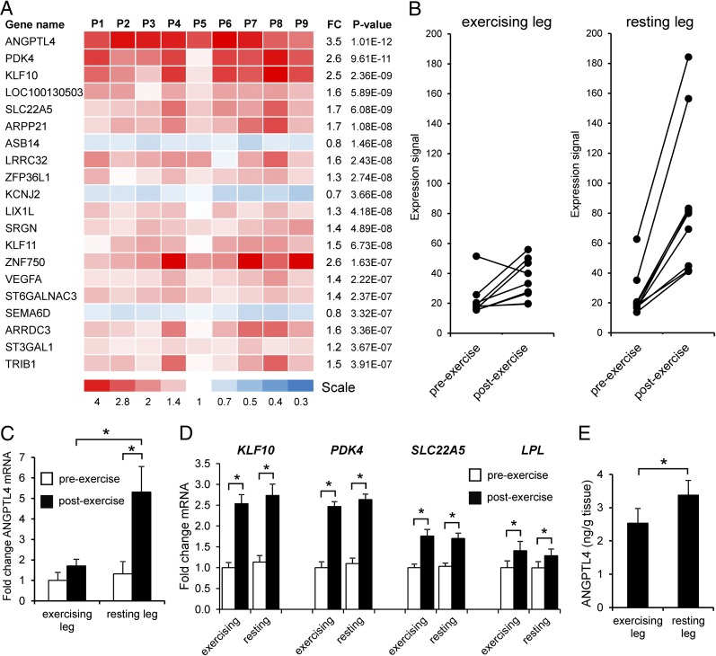
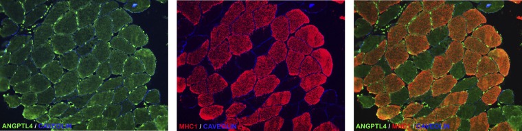
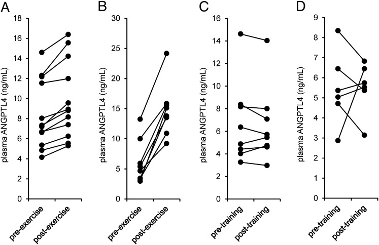
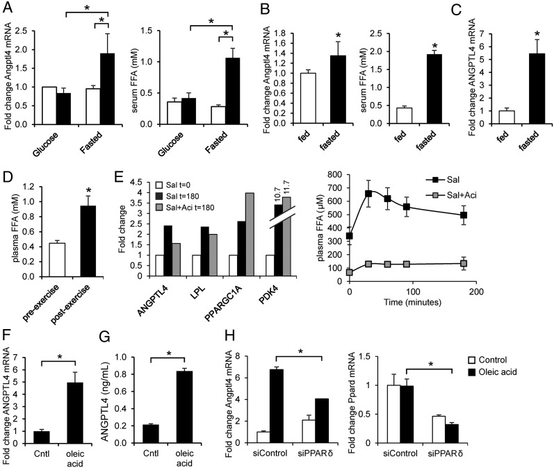
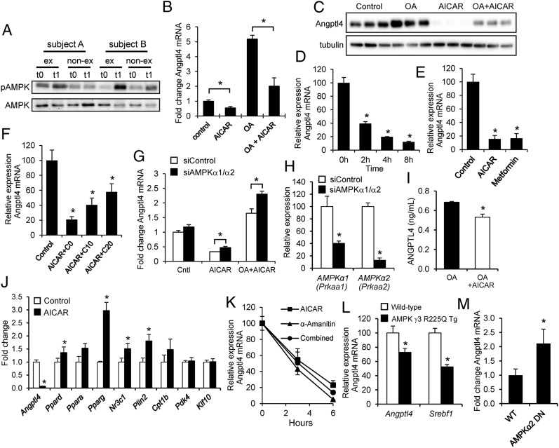
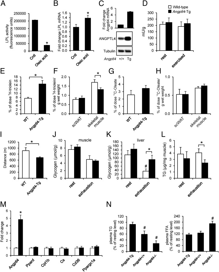
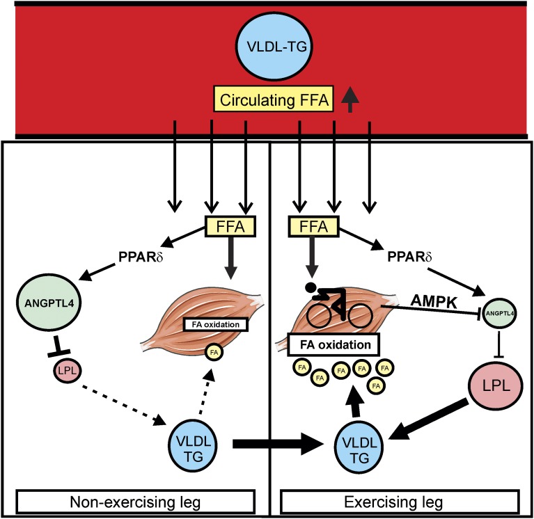
Physical activity increases energy metabolism in exercising muscle. Whether acute exercise elicits metabolic changes in nonexercising muscles remains unclear. We show that one of the few genes that is more highly induced in nonexercising muscle than in exercising human muscle during acute exercise encodes angiopoietin-like 4 (ANGPTL4), an inhibitor of lipoprotein lipase-mediated plasma triglyceride clearance. Using a combination of human, animal, and in vitro data, we show that induction of ANGPTL4 in nonexercising muscle is mediated by elevated plasma free fatty acids via peroxisome proliferator-activated receptor-δ, presumably leading to reduced local uptake of plasma triglyceride-derived fatty acids and their sparing for use by exercising muscle. In contrast, the induction of ANGPTL4 in exercising muscle likely is counteracted via AMP-activated protein kinase (AMPK)-mediated down-regulation, promoting the use of plasma triglycerides as fuel for active muscles. Our data suggest that nonexercising muscle and the local regulation of ANGPTL4 via AMPK and free fatty acids have key roles in governing lipid homeostasis during exercise.


Conflict of interest statement

The authors declare no conflict of interest.

Figures

Fig. 1.
Fig. 1.
Exercise induces ANGPTL4 gene expression in nonexercising human muscle. (A) Heatmap showing genes altered by exercise in the nonexercising leg muscle ranked by statistical significance. Values are displayed per subject (P1 to P9). Fold-change (FC) in gene expression is indicated. (B) mRNA expression profile of ANGPTL4 in the exercising and nonexercising legs according to microarray. (C) qPCR analysis of ANGPTL4 mRNA expression. (D) mRNA expression of PPARδ targets KLF10, PDK4, and SLC22A5, as well as LPL. (E) ANGPTL4 protein levels in postexercise muscle biopsies as determined by ELISA. Error bars represent SEM. *Significantly different according to paired Student t test (P < 0.01).
Fig. 2.
Fig. 2.
ANGPTL4 protein is detected in human muscle in myocytes and endothelial cells. Representative images of immunofluorescent staining of ANGPTL4 in a biopsy from human vastus lateralis muscle show ANGPTL4 in green, myosin heavy chain 1 (MHC1), a marker type I fiber, in red, and caveolin in blue.
Fig. 3.
Fig. 3.
Plasma ANGPTL4 levels are increased by acute exercise but not by exercise training. (A) Plasma ANGPTL4 levels before and after 1 h of one-legged cycling exercise at 50% of the one-legged Wmax (study A, n = 12). (B) Plasma ANGPTL4 levels before and after 3 h of cycling exercise at 40% Wmax (Study B, n = 8). (C) Fasting plasma ANGPTL4 levels before and after an intense 2-wk endurance training program on a cycling ergometer (study C, n = 8). (D) Fasting plasma ANGPTL4 levels before and after a moderate-intensity, 12-wk endurance training program on a cycling ergometer (study D, n = 6).
Fig. 4.
Fig. 4.
Sensitive induction of the ANGPTL4 gene by FFAs in human and mouse myocytes. (A) C2C12 myotubes were incubated for 6 h with 10% serum from subjects (n = 5) before exercise (white bar) and after exercise (black bar) performed in fasted state or with provision of glucose (study E). (Left) Angptl4 mRNA. (Right) Serum FFA levels. (B) C2C12 myotubes were incubated for 3 h with 10% serum from subjects (n = 12) at the end of a 60-h fast or after 60 h in the normal fed condition (study F). (Left) Angptl4 mRNA levels. (Right) Serum FFA levels. (C) ANGPTL4 mRNA in muscle biopsies collected at the end of the 60-h fast or after 60 h in the normal fed condition (study F). (D) Plasma FFA levels before and after one-legged exercise (n = 12). (E) (Left) Pooled mRNA expression of selected genes in muscle biopsies collected before and after salbutamol infusion with and without prior acipimox administration (study G, n = 9). (Right) Plasma FFA levels during salbutamol (Sal) infusion. Error bars represent SEM. (F and G) ANGPTL4 mRNA (F) and ANGPTL4 (G) concentration in medium in primary human myotubes treated with oleic acid. (H) Angptl4 and Ppard mRNA in C2C12 myotubes transfected with control (nontargeting) or with PPARδ siRNA and treated with oleic acid. *Significantly different according to Student t test (P < 0.05). Error bars represent SD unless otherwise indicated. Cells were treated for 12 h unless otherwise indicated.
Fig. 5.
Fig. 5.
AMPK activation suppresses Angptl4 mRNA. (A) Immunoblot for AMPK and phospho-AMPK in skeletal muscle biopsies from two selected subjects before (t0) and after (t1) exercise. (B) Expression of Angptl4 mRNA in C2C12 myotubes treated with oleic acid (200 μM) and/or AICAR (1 mM) for 3 h. (C) Immunoblot for ANGPTL4 in C2C12 myotubes treated with oleic acid and/or AICAR. (D) Time-course of the effect of AICAR on Angptl4 mRNA in C2C12 myotubes. (E) Comparison of the effect of AICAR (1 mM) and metformin (0.5 mM) on Angptl4 mRNA in C2C12 myotubes. (F) Effect of AICAR (1 mM) and compound C cotreatment on Angptl4 mRNA in C2C12 myotubes. Concentrations are indicated in millimolars. (G) Angptl4 mRNA in C2C12 myotubes transfected with control (nontargeting) or AMPKα1/AMPKα2 siRNA and treated with AICAR. (H) Effective knockdown of AMPKα1 and AMPKα2 by AMPKα1/AMPKα2 siRNA. (I) ANGPTL4 levels in medium of human primary myotubes treated with oleic acid and AICAR. (J) Expression of PPARs and PPAR targets in C2C12 myotubes treated with AICAR. (K) Angptl4 mRNA in C2C12 myotubes preincubated with 50 μg/mL α-Amanitin for 1 h and treated with AICAR for 3 h or 6 h. (L) Angptl4 mRNA in the gastrocnemius of mice that overexpress an activating mutant of the muscle-specific isoform of the AMPKγ subunit. Error bars represent SEM. Data were extracted from GSE4065 (22). (M) Angptl4 mRNA in the gastrocnemius of mice that overexpress a dominant-negative mutant of the AMPKα2 subunit. Cells were treated for 12 h unless otherwise indicated. Error bars represent SEM. *Significantly different according to Student t test (P < 0.05). Error bars represent SD unless otherwise indicated.
Fig. 6.
Fig. 6.
Angptl4 up-regulation impairs LPL activity and uptake of plasma TG-derived fatty acids in muscle. (A and B) Heparin-releasable LPL activity (A) and Lpl mRNA (B) in mouse C2C12 myotubes treated with oleic acid (400 µM) for 24 h. (C) ANGPTL4 protein abundance and Angptl4 mRNA expression in mouse skeletal muscle. (D) Total LPL activity in skeletal muscle (gastrocnemius) of WT and Angptl4-Tg mice at rest and after 90 min of moderate running exercise (12 m/min). (E) Serum 3H activity after 15 min of running in WT and Angptl4-Tg mice injected with [14C]-oleate together with glycerol tri[3H]oleate-labeled VLDL-like particles. (F) 3H-activity in subcutaneous adipose tissue and gastrocnemius after 15 min of running. (G) Serum 14C activity after 15 min of running in WT and Angptl4-Tg mice injected with [14C]-oleate together with glycerol tri[3H]oleate-labeled VLDL-like particles. (H) 14C activity in subcutaneous adipose tissue and gastrocnemius after 15 min of running. (I) Distance covered, excluding warm-up, by WT and Angptl4-Tg mice subjected to an incremental exercise test to exhaustion. (JL) Muscle glycogen (J), liver glycogen (K), and muscle TG (L) levels in WT and Angptl4-Tg mice at rest or after exhaustive running exercise. (M) mRNA expression of selected genes in skeletal muscle (gastrocnemius) of WT and Angptl4-Tg mice at rest. (N) The relative levels of plasma TG (Left) and FFA (Right) in the exercised state (90 min of moderate running exercise) compared with the resting state (90 min rest) in WT, Angptl4-Tg, and Angptl4−/− mice. *Significantly different from WT mice according to Student t test (P < 0.05). #Significantly different from resting mice according to Student t test (P < 0.05). Error bars represent SEM; n = 6–10 mice per group.
Fig. 7.
Fig. 7.
Schematic representation of the proposed role of ANGPTL4 in providing lipid to exercising muscle. During exercise, circulating FFAs and VLDL particles are directed to exercising and nonexercising muscle. In the nonexercising leg, increased FFA levels provoke an increase in ANGPTL4 expression via PPARδ, leading to inhibition of LPL activity and consequent reduction in uptake of fatty acids derived from VLDL, which likely is aimed at preventing lipid overload. In contrast, in the exercising leg the stimulatory effect of FFA on ANGPTL4 mRNA is counteracted by AMPK-mediated suppression of ANGPTL4 mRNA. As a result, LPL activity remains high, allowing full exploitation of fatty acids derived from VLDL as the substrate for fatty acid oxidation to meet the energetic needs of exercising muscle.

References

    1. Kiens B. Skeletal muscle lipid metabolism in exercise and insulin resistance. Physiol Rev. 2006;86(1):205–243. - PubMed
    1. Keller P, et al. Using systems biology to define the essential biological networks responsible for adaptation to endurance exercise training. Biochem Soc Trans. 2007;35(Pt 5):1306–1309. - PubMed
    1. Buford TW, Cooke MB, Willoughby DS. Resistance exercise-induced changes of inflammatory gene expression within human skeletal muscle. Eur J Appl Physiol. 2009;107(4):463–471. - PubMed
    1. Catoire M, et al. Pronounced effects of acute endurance exercise on gene expression in resting and exercising human skeletal muscle. PLoS ONE. 2012;7(11):e51066. - PMC - PubMed
    1. Schmutz S, et al. Endurance training modulates the muscular transcriptome response to acute exercise. Pflugers Arch. 2006;451(5):678–687. - PubMed

Publication types

Associated data