. 2014 Mar 25;111(12):4590-5.
doi: 10.1073/pnas.1315275111. Epub 2014 Mar 3.

Categorical encoding of color in the brain

Affiliations

Categorical encoding of color in the brain

Chris M Bird et al. Proc Natl Acad Sci U S A. .

Abstract

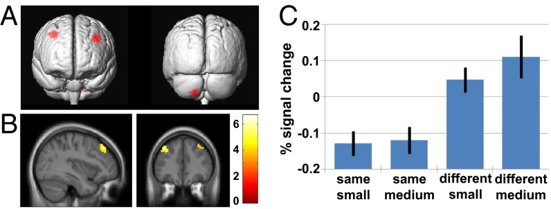
The areas of the brain that encode color categorically have not yet been reliably identified. Here, we used functional MRI adaptation to identify neuronal populations that represent color categories irrespective of metric differences in color. Two colors were successively presented within a block of trials. The two colors were either from the same or different categories (e.g., "blue 1 and blue 2" or "blue 1 and green 1"), and the size of the hue difference was varied. Participants performed a target detection task unrelated to the difference in color. In the middle frontal gyrus of both hemispheres and to a lesser extent, the cerebellum, blood-oxygen level-dependent response was greater for colors from different categories relative to colors from the same category. Importantly, activation in these regions was not modulated by the size of the hue difference, suggesting that neurons in these regions represent color categorically, regardless of metric color difference. Representational similarity analyses, which investigated the similarity of the pattern of activity across local groups of voxels, identified other regions of the brain (including the visual cortex), which responded to metric but not categorical color differences. Therefore, categorical and metric hue differences appear to be coded in qualitatively different ways and in different brain regions. These findings have implications for the long-standing debate on the origin and nature of color categories, and also further our understanding of how color is processed by the brain.

Keywords: categorization; chromatic; functional magnetic resonance imaging.


Conflict of interest statement

The authors declare no conflict of interest.

Figures

Fig. 1.
Fig. 1.
fMRI adaptation stimuli and design. (A) Flow and time course of blocked design. During color stimulation a colored square was presented centrally on gray background (400 ms), 12 times separated by a gray background for 400 ms. (B) Two colors were presented six times each within a color stimulation block. On 12.5% blocks, one of the colored stimuli had a target that was a lighter square presented in the center of the colored stimulus (see right side of B). Participants were required to press a key when the target was detected, and blocks with targets were excluded from the analysis. (C) Print-rendered versions of the colors used. The dashed line indicates the blue-green lexical distinction made by the majority of the participants (n = 17): one stimulus was named green (G1) and three were named blue (B1/B2/B3). (D) Table of conditions. Across runs, all colors were paired with every other color, including itself, giving blocks where colors were either the same- or different-category, and where the size of the hue difference was absent (identical), small, medium, or large. The red box indicates the 2 × 2 design used in one analysis which aimed to identify regions of the brain that respond to categorical but not metric changes in color.
Fig. 2.
Fig. 2.
Results of univariate analysis of the main effect of color category. (A) Main effect of color category in left and right MFG and cerebellum projected on to the group averaged structural scan (P < 0.001 uncorrected for multiple comparisons, extent threshold = 20 contiguous voxels). (B) The same contrast shown centered on the peak voxel in the left MFG. The colorbar represents the value of the t-statistic. (C) Mean percentage signal change for the four experimental block types compared with baseline shown for the peak voxel in the left MFG (error bars show SEMs). The baseline is an unconstrained “rest” period, and therefore it is the differences between experimental conditions that are critical rather than the absolute change relative to baseline.
Fig. 3.
Fig. 3.
RSA analyses of experimental blocks and color metric differences. Whole-brain “searchlight” analyses were carried out where the similarity in the pattern BOLD signal across 257 voxels in a moving sphere were compared against different phases of the experiment. The voxel at the center of the sphere was assigned the t-statistic for the comparison. (A) Brain regions where the similarity in patterns of BOLD signal was higher during experimental blocks than during IBIs, centered on the peak voxel showing the effect (P < 0.001 uncorrected). The colorbar represents the value of the t-statistic. (B) Bar graph showing the mean correlations between the experimental blocks and the interbloc intervals for the peak voxel identified in A, averaged across all participants (error bars show SEMs). Note that this is for illustrative purposes and does not represent an independent analysis. (C) Brain regions where the similarity in patterns of activations was greater during blocks with small rather than medium hue difference (color metric effect), centered on the peak voxel showing the effect (P < 0.001 uncorrected). The colored bar represents the value of the t-statistic. (D) Bar graph showing the mean correlations between blocks with small hue difference versus small and medium hue difference (error bars show SEMs). Note that this is for illustrative purposes and does not represent an independent analysis.

References

    1. Regier T, Kay P. Language, thought, and color: Whorf was half right. Trends Cogn Sci. 2009;13(10):439–446. - PubMed
    1. Regier T, Kay P, Cook RS. Focal colors are universal after all. Proc Natl Acad Sci USA. 2005;102(23):8386–8391. - PMC - PubMed
    1. Franklin A, Davies IRL. New evidence for infant color categories. Br J Dev Psychol. 2004;22:349–377.
    1. Roberson D, Davies IRL, Davidoff J. Color categories are not universal: Replications and new evidence from a stone-age culture. J Exp Psychol Gen. 2000;129(3):369–398. - PubMed
    1. Regier T, Kay P, Khetarpal N. Color naming reflects optimal partitions of color space. Proc Natl Acad Sci USA. 2007;104(4):1436–1441. - PMC - PubMed

Publication types

LinkOut - more resources