. 2014 Mar 18;111(11):4145-50.
doi: 10.1073/pnas.1320715111. Epub 2014 Mar 3.

Seminal vesicle protein SVS2 is required for sperm survival in the uterus

Affiliations

Seminal vesicle protein SVS2 is required for sperm survival in the uterus

Natsuko Kawano et al. Proc Natl Acad Sci U S A. .

Abstract

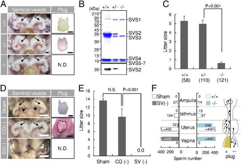
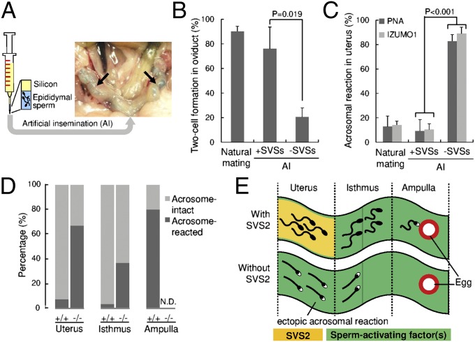
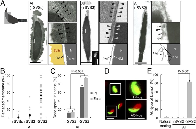
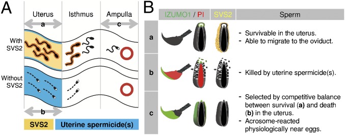
In mammals, sperm migrate through the female reproductive tract to reach the egg; however, our understanding of this journey is highly limited. To shed light on this process, we focused on defining the functions of seminal vesicle secretion 2 (SVS2). SVS2(-/-) male mice produced sperm but were severely subfertile, and formation of a copulatory plug to cover the female genital opening did not occur. Surprisingly, even when artificial insemination was performed with silicon as a substitute for the plug, sperm fertility in the absence of SVS2 remained severely reduced because the sperm were already dead in the uterus. Thus, our results provide evidence that the uterus induces sperm cell death and that SVS2 protects sperm from uterine attack.

Keywords: acrosomal reaction; decapacitation; in vivo fertilization; uterine sperm selection; uterine spermicide.


Conflict of interest statement

The authors declare no conflict of interest.

Figures

Fig. 1.
Fig. 1.
Impaired copulatory plug formation in SVS2−/− and seminal vesicle-excised mice. (A) Appearance of seminal vesicles and copulatory plugs. Arrows, seminal vesicles. N.D., not detected. (Scale bars, 2 mm.) (B) Immunoblotting of the total inner fluid of seminal vesicles isolated from male mice with anti-SVS2 pAb. (Upper) Staining with CBB; (Lower) reaction with anti-SVS2 pAb. (C) Fecundity of male mice. Parentheses, numbers of male mice examined. (D) Appearance of seminal vesicles and copulatory plugs in surgically operated mice. Sham, sham-operated mice; CG(-), mice with coagulating glands excised; SV(-), mice with seminal vesicles excised. Arrows, seminal vesicles; arrowheads, coagulating glands; dotted circle, a representative plug formed by a CG(-) male mouse. (Scale bars, 2 mm.) (E) Fecundity of surgically operated male mice (n = 5). N.S., not significant. (F) Numbers of sperm entering the female reproductive tract after copulation. The total sperm number is indicated as the sum of five independent experiments. (Right graph) Number of sperm detected in each sectioned tract of female mice mated with SVS2+/+ or SVS2−/− male mice. (Left graph) Number of sperm detected in each sectioned tract of female mice mated with Sham or SV(-) male mice.
Fig. 2.
Fig. 2.
Sperm fertility with or without SVS2 by AI using silicon as a substitute for the copulatory plug. (A) Experimental design of the AI procedure. Arrows, sperm solution coinjected with a blue dye in the uterine cavity. (B) Formation of two-cell embryos in oviducts by injection of sperm with or without SVSs using AI. Concomitantly, when female mice were mated with male mice, the number of two-cell embryos formed in oviducts was counted (n = 6). (C) Rates of acrosome-reacted sperm in the uterus by injection of sperm with or without SVSs using AI. Concomitantly, when female mice were mated with male mice, the number of acrosome-reacted sperm in the uterus was counted (n = 3). (D) Rates of acrosome-reacted sperm ejaculated from SVS2−/− mice or control mice (SVS2+/+and SVS2+/−) to the female reproductive tract (n = 3). In three parts of the reproductive tract, the number of acrosome-reacted sperm (RFP-positive and GFP-negative) was counted and compared with that of acrosome-intact sperm (RFP-positive and GFP-positive). (E) Schematic model of ectopic sperm activation in the absence of SVS2. Sperm with black heads, acrosome-intact sperm; sperm with white heads, acrosome-reacted sperm. Green substance, predicted sperm-activating factor(s).
Fig. 3.
Fig. 3.
SVS2-mediated protection of intrauterine sperm from membrane disruption. (A) Transmission electron microscopic (TEM) images. Diagram at far left indicates the orientation of sectioned heads of sperm. (Left) Sperm with SVSs; (Center) sperm with SVS2; (Right) sperm without SVS2. (Left, Center, and Right) Right upper and middle images are enlarged views of boxes in the left images, and diagrams are depicted in the right lower images. PM, plasma membrane; N, nucleus; NM, nuclear membrane. (Scale bars: 1 μm.) (Center, Inset) A fluorescent image of the sperm reacted with anti-SVS2 pAb. (Scale bar, 5 μm.) (B) Rates of broken membrane areas in TEM images (n = 20). *P < 0.001. (C) Rates of dead sperm in the uterus determined by staining with PI and eosin (n = 3). (D) Fluorescent images of sperm collected from the uterus by staining with anti-IZUMO1 mAb (green) and DAPI (red). (Right) Enlarged images of boxes at Left. H-type, fusion-capable sperm; AC-type, dead sperm. (Scale bars, 10 μm.) (E) Rate of sperm categorized as AC-type (n = 3).
Fig. 4.
Fig. 4.
Schematic model of the role of SVS2 in sperm protection in vivo. (A) Sperm migration in the female reproductive tract. Sperm with black heads, acrosome-intact sperm; sperm with white heads, acrosome-reacted sperm. Yellow, SVS2; blue, predicted maternal spermicide(s). a, acrosome-intact sperm penetrated the uterus after natural mating. b, membrane-disrupted dead sperm in the uterus in the absence of SVS2. c, acrosome-reacted sperm penetrated the ampulla. (B) Status of acrosomal and plasma membranes of sperm in the female reproductive tract. Categories a, b, and c correspond to those in A. Green, sperm membrane stained with anti-IZUMO1 mAb; red, sperm nucleus stained with PI.

References

    1. Suarez SS (2006) in Knobil and Neill’s Physiology of Reproduction, ed Neill JD (Academic, New York), 3rd Ed, pp 113–145.
    1. Chang MC. A detrimental effect of seminal plasma on the fertilizing capacity of sperm. Nature. 1957;179(4553):258–259. - PubMed
    1. Bedford JM, Chang MC. Removal of decapacitation factor from seminal plasma by high-speed centrifugation. Am J Physiol. 1962;202(1):179–181. - PubMed
    1. Fraser LR. Mouse sperm capacitation in vitro involves loss of a surface-associated inhibitory component. J Reprod Fertil. 1984;72(2):373–384. - PubMed
    1. Nixon B, et al. The identification of mouse sperm-surface-associated proteins and characterization of their ability to act as decapacitation factors. Biol Reprod. 2006;74(2):275–287. - PubMed

Publication types

MeSH terms