. 2014 Mar 18;111(11):4221-6.
doi: 10.1073/pnas.1320753111. Epub 2014 Mar 3.

Circulating hematopoietic stem and progenitor cells are myeloid-biased in cancer patients

Affiliations

Circulating hematopoietic stem and progenitor cells are myeloid-biased in cancer patients

Wen-Chao Wu et al. Proc Natl Acad Sci U S A. .

Abstract

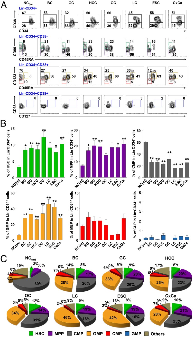
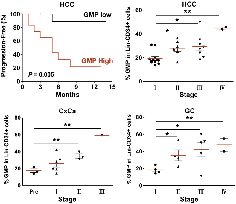
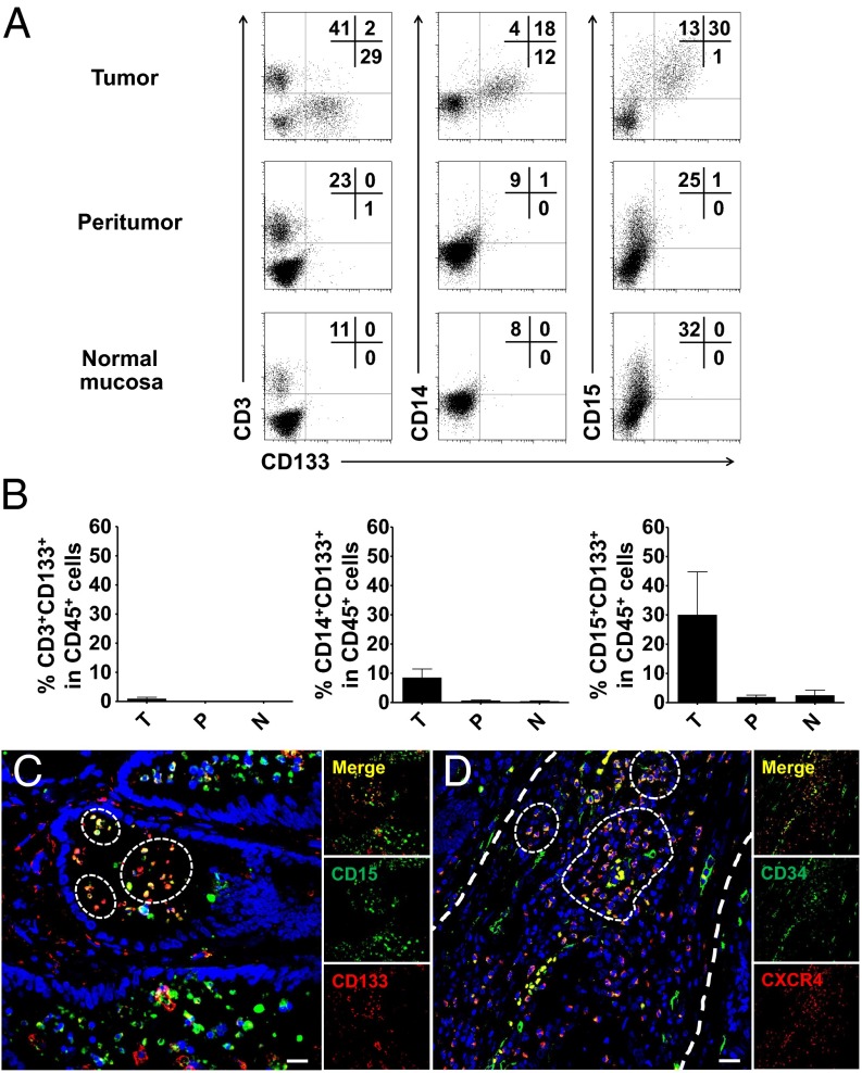
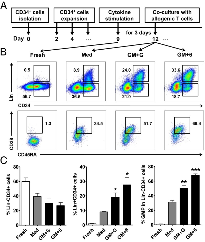
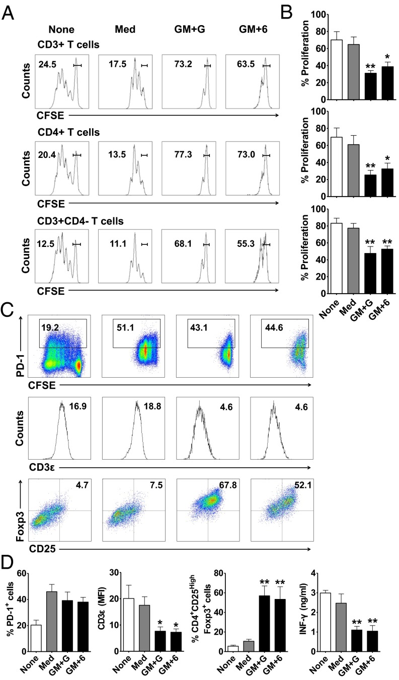
Cancer is associated with a profound perturbation in myelopoiesis that results in the accumulation of myeloid-derived suppressor cells (MDSCs) to promote disease progression. Recent studies in mice suggest that tumor-derived factors could regulate the differentiation of hematopoietic stem and progenitor cells (HSPCs) in the bone marrow and subsequently contribute to dysregulation of hematopoiesis. However, the nature and role of HPSCs in patients with cancer remain unknown. Here we show, in detailed studies of the peripheral blood from 133 untreated patients with seven different types of tumors, that the composition of circulating HSPCs was significantly altered in patients with solid tumors. The frequencies of circulating granulocyte-monocyte progenitors (GMPs) were increased four to seven fold in all types of tumors examined, and the circulating hematopoietic precursors exhibited myeloid bias with a skew toward granulocytic differentiation in patients with solid tumors. These myeloid precursors are selectively enriched in tumor tissues, and the high levels of circulating GMPs were positively correlated with disease progression. By using cord blood-derived CD34(+) cells, we developed an in vitro short-term culture model to effectively induce the rapid generation of MDSCs. We found that, among the factors produced by various tumors, GM-CSF, granulocyte colony-stimulating factor, and IL-6 could not only promote the myeloid-biased differentiation, but also induce the differentiation of myeloid precursors into functional MDSCs. These findings suggest that the altered circulating HSPCs may serve as an important link between dysregulated bone marrow hematopoiesis and accumulated MDSCs in patients with cancer.


Conflict of interest statement

The authors declare no conflict of interest.

Figures

Fig. 1.
Fig. 1.
Altered composition of circulating HSPC subsets with a skew toward GMPs in patients with cancer. (A) Representative FACS staining of HSPC subsets in the peripheral blood LinCD34+ cells from healthy donors and patients with cancer. HCC (n = 26); and BC, breast cancer (n = 9); CxCa, cervical cancer (n = 19); ESC, esophageal cancer (n = 4); GC, gastrointestinal cancer (n = 18); LC, lung cancer (n = 8); NC, age-matched healthy donors (n = 63); OC, ovarian cancer (n = 4). (B) Summary of the circulating HSPC subsets in LinCD34+ population. Statistical differences between groups were calculated by one-way ANOVA, followed by post hoc Bonferroni tests (*P < 0.05 and **P < 0.01 vs. healthy donors). (C) Pie charts summarizing the frequencies of HSPC subsets from healthy donors and patients.
Fig. 2.
Fig. 2.
Increased circulating GMPs predicted poor survival in patients with cancer. Twenty-eight patients with HCC were divided into two groups according to the median value of GMP frequency. Individuals in the low GMP group (black) had significantly improved survival compared with those in the high GMP group (red). The time to progression was estimated by Kaplan–Meier method and compared by using the log-rank test. The frequency of GMPs in the LinCD34+ cells from patients with HCC (Upper Right), cervical cancer (CxCa, Lower Left), and gastrointestinal cancer (GC, Lower Right) are shown separated by their clinical stages (*P < 0.05 and **P < 0.01).
Fig. 3.
Fig. 3.
IMCs are enriched in colon tumor tissues. (A) Representative FACS analysis of CD133+CD3+, CD133+CD14+, and CD133+CD15+ cells in tissue CD45+ cells isolated from eight paired colon tumor, peritumor, and relative normal mucosal tissues. (B) Summary of the results from A. (C) Multiple staining of CD133 (red), CD15 (green), and DAPI (blue) in frozen sections were analyzed by confocal microscopy. The coexistence of CD133 and CD15 confirmed that a proportion of CD133+CD15+ cells in colon tumor tissues. One of 10 representative micrographs is shown. (D) Multiple staining of CXCR4 (red), CD34 (green), and DAPI (blue) in paraffin-embedded sections shows the coexistence of CXCR4- and CD34-positive cell in tumor tissues. One of 10 representative micrographs is shown. (Scale bar, 20 μm.)
Fig. 4.
Fig. 4.
GM-CSF, G-CSF, and IL-6 promote the differentiation of GMP and Lin+CD34+ cells. (A) Freshly isolated CD34+ cells from CB mononuclear cells were cultured in HSC expansion media for 8–10 d. The expanded cells were then stimulated with cytokines for 3 d, followed by coculture with allogeneic T cells. (B) Representative flow cytometry data of LinCD34+ cells, Lin+CD34+ cells, and GMPs after stimulation. (C) Summary of the frequency of LinCD34+ and Lin+CD34+ cells in total nucleated cells and of the GMPs in LinCD34+ cells after stimulation. The values given represent the mean (±SE) of four separate experiments; *P < 0.05, **P < 0.01, and ***P < 0.001 vs. untreated expanded cells (Med).
Fig. 5.
Fig. 5.
CB-MDSCs exhibit strong suppressive activity against T cells, down-regulate CD3ε expression, and induce Foxp3+ regulatory T cells. (A) Carboxyfluorescein diacetate succinimidyl ester-labeled pan-T cells were cocultured at a 1:1 ratio with or without CB-MDSCs for 6 d, and then analyzed by FACS. The percentages of undivided cells are shown. (B) Summary of the proliferation of T cells. (C) Representative staining of PD-1, CD3ε, and Foxp3 expression on T cells. (D) Summary of the frequency of PD-1+ and CD25+Foxp3+ and the mean fluorescence intensity of CD3ε expression on T cells. IFN-γ concentrations were determined from culture supernatants. The data shown represent the mean (±SE) of four separate experiments; *P < 0.05 and **P < 0.01 vs. pan-T cells cocultured with untreated expanded cells (Med).

References

    1. Kondo M, et al. Biology of hematopoietic stem cells and progenitors: implications for clinical application. Annu Rev Immunol. 2003;21:759–806. - PubMed
    1. Laiosa CV, Stadtfeld M, Graf T. Determinants of lymphoid-myeloid lineage diversification. Annu Rev Immunol. 2006;24:705–738. - PubMed
    1. Essers MA, et al. IFNalpha activates dormant haematopoietic stem cells in vivo. Nature. 2009;458(7240):904–908. - PubMed
    1. King KY, Goodell MA. Inflammatory modulation of HSCs: Viewing the HSC as a foundation for the immune response. Nat Rev Immunol. 2011;11(10):685–692. - PMC - PubMed
    1. Doulatov S, Notta F, Laurenti E, Dick JE. Hematopoiesis: A human perspective. Cell Stem Cell. 2012;10(2):120–136. - PubMed

Publication types

MeSH terms

Substances