

Three-dimensional ultrasound as a predictor of pregnancy in patients undergoing ART
- PMID: 24592022
- PMCID: PMC3939135
- DOI: 10.5152/jtgga.2012.15
Three-dimensional ultrasound as a predictor of pregnancy in patients undergoing ART
Abstract
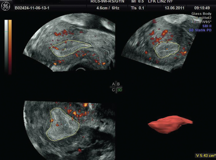
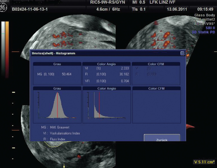
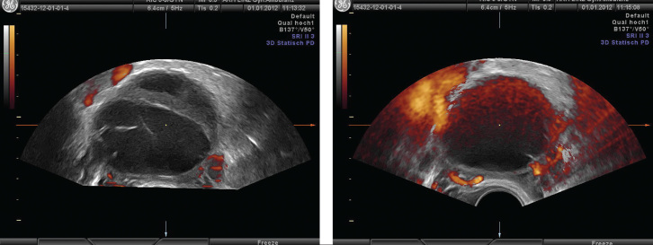
Different ultrasound parameters have been used to assess endometrial receptivity during ART treatment, including endometrial thickness, endometrial pattern, endometrial volume, Doppler of uterine arteries and endometrial blood flow. However, conflicting results have been reported with regard to their role in the prediction of pregnancy in ART treatment. The 3D ultrasound with power Doppler provides a unique tool with which to examine the blood supply of the whole endometrium and subendometrial region. Volume assessment can also be precisely performed by 3D ultrasound. Based on a med-line research and on our experience, the clinical use of 3D ultrasound is discussed in this review article.
ART tedavisi sırasında endometriyumun kabul ediciliğini değerlendirmek için farklı ultrason parametreleri kullanılmaktadır. Bunlar içinde endometriyal kalınlık, endometriyal patern, endometriyal hacim, uterus arterlerinin ve endometriyal kan akımının Doppler’i yer almaktadır. Bununla beraber, ART tedavisinde gebeliğin öngörülmesindeki rolleri ile ilgili olarak birbiriyle çelişen sonuçlar bildirilmiştir. Bütün endometriyumun ve subendometriyal bölgenin kan akımının incelenmesinin mümkün olduğu power Doppler’li 3D ultrason yegane bir araç sağlamaktadır. 3D ultrason ile hacim değerlendirmeleri de kesin olarak yapılabilmektedir. Med-line araştırması ve kendi deneyimimize dayanarak, bu derleme makalede 3D ultrasonun klinik kullanımı tartışılmaktadır.
Keywords: 3D; ART; IVF; power Doppler; ultrasound.
Figures
Similar articles
-
The role of endometrial and subendometrial blood flows measured by three-dimensional power Doppler ultrasound in the prediction of pregnancy during IVF treatment.Hum Reprod. 2006 Jan;21(1):164-70. doi: 10.1093/humrep/dei277. Epub 2005 Aug 25. Hum Reprod. 2006. PMID: 16123083
-
The role of endometrial and subendometrial vascularity measured by three-dimensional power Doppler ultrasound in the prediction of pregnancy during frozen-thawed embryo transfer cycles.Hum Reprod. 2006 Jun;21(6):1612-7. doi: 10.1093/humrep/dei502. Epub 2006 Jan 31. Hum Reprod. 2006. PMID: 16449309
-
The role of endometrial blood flow measured by three-dimensional power Doppler ultrasound in the prediction of pregnancy during in vitro fertilization treatment.Eur J Obstet Gynecol Reprod Biol. 2007 Nov;135(1):8-16. doi: 10.1016/j.ejogrb.2007.06.006. Epub 2007 Jul 20. Eur J Obstet Gynecol Reprod Biol. 2007. PMID: 17658677 Review.
-
Endometrial and subendometrial vascularity are significantly lower in patients with endometrial volume 2.5 ml or less.Reprod Biomed Online. 2009 Feb;18(2):262-8. doi: 10.1016/s1472-6483(10)60264-7. Reprod Biomed Online. 2009. PMID: 19192348
-
Ultrasound in assisted reproduction: a call to fill the endometrial gap.Fertil Steril. 2016 Jun;105(6):1394-1402.e4. doi: 10.1016/j.fertnstert.2016.04.012. Epub 2016 Apr 29. Fertil Steril. 2016. PMID: 27140291 Review.
Cited by
-
Age-Related Dynamics in Endometrial Vascularity: A Comprehensive Three-Dimensional Ultrasound Evaluation During Follicular and Luteal Phases.J Clin Med. 2025 Jun 18;14(12):4332. doi: 10.3390/jcm14124332. J Clin Med. 2025. PMID: 40566077 Free PMC article.
-
Automated endometrial identification and volume calculation in normal uteri using a novel smart ERA technique.Sci Rep. 2024 Sep 4;14(1):20525. doi: 10.1038/s41598-024-71069-z. Sci Rep. 2024. PMID: 39227624 Free PMC article.
-
The role of color Doppler in assisted reproduction: A narrative review.Int J Reprod Biomed. 2019 Dec 26;17(11):779-788. doi: 10.18502/ijrm.v17i10.5484. eCollection 2019 Dec. Int J Reprod Biomed. 2019. PMID: 31911960 Free PMC article. Review.
-
Acupuncture in improving endometrial receptivity: a systematic review and meta-analysis.BMC Complement Altern Med. 2019 Mar 13;19(1):61. doi: 10.1186/s12906-019-2472-1. BMC Complement Altern Med. 2019. PMID: 30866920 Free PMC article.
-
Letrozole Versus Clomiphene Citrate and Natural Cycle: Endometrial Receptivity During Implantation Window in Women With Polycystic Ovary Syndrome.Front Endocrinol (Lausanne). 2021 Jan 18;11:532692. doi: 10.3389/fendo.2020.532692. eCollection 2020. Front Endocrinol (Lausanne). 2021. PMID: 33537000 Free PMC article. Clinical Trial.
References
-
- Jurkovic D, Geipel A, Gruboeck K, Jauniaux E, Natucci M, Campbell S. Three-dimensional ultrasound for the assessment of uterine anatomy and detection of congenital anomalies: a comparison with hysterosalpingography and two-dimensional sonography. Ultrasound Obstet Gynecol. 1995;5:233–7. - PubMed
-
- Weismann CF, Datz L. Diagnostic algorithm: how to make use of new 2D, 3D and 4D ultrasound technologies in breast imaging. Eur J Radiol. 2007;64:250–7. - PubMed
-
- Yaman C, Fridrik M. Three-dimensional ultrasound to assess the response to treatment in gynecological malignancies. Gynecol Oncol. 2005;97:665–8. - PubMed
-
- Yaman C, Ebner T, Jesacher K. Three-dimensional power Doppler in the diagnosis of ovarian torsion. Ultrasound Obstet Gynecol. 2002;20:513–5. - PubMed
-
- Yaman C, Habelsberger A, Tews G, Pölz W, Ebner T. The role of three-dimensional volume measurement in diagnosing endometrial cancer in patients with postmenopausal bleeding. Gynecol Oncol. 2008;110:390–5. - PubMed
Publication types
LinkOut - more resources
Full Text Sources