

Nanocomposite scaffold for chondrocyte growth and cartilage tissue engineering: effects of carbon nanotube surface functionalization
- PMID: 24593020
- PMCID: PMC4172384
- DOI: 10.1089/ten.TEA.2013.0328
Nanocomposite scaffold for chondrocyte growth and cartilage tissue engineering: effects of carbon nanotube surface functionalization
Abstract
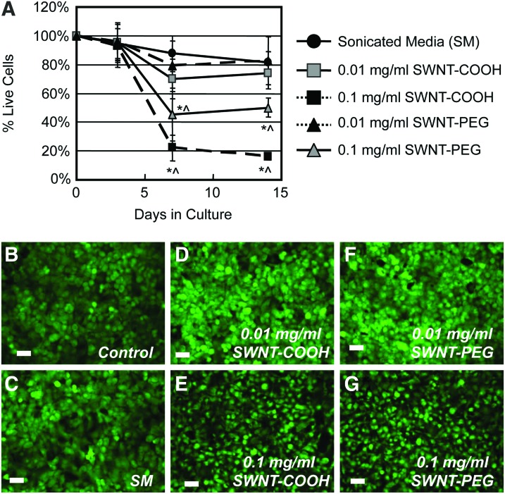
The goal of this study was to assess the long-term biocompatibility of single-wall carbon nanotubes (SWNTs) for tissue engineering of articular cartilage. We hypothesized that SWNT nanocomposite scaffolds in cartilage tissue engineering can provide an improved molecular-sized substrate for stimulation of chondrocyte growth, as well as structural reinforcement of the scaffold's mechanical properties. The effect of SWNT surface functionalization (-COOH or -PEG) on chondrocyte viability and biochemical matrix deposition was examined in two-dimensional cultures, in three-dimensional (3D) pellet cultures, and in a 3D nanocomposite scaffold consisting of hydrogels+SWNTs. Outcome measures included cell viability, histological and SEM evaluation, GAG biochemical content, compressive and tensile biomechanical properties, and gene expression quantification, including extracellular matrix (ECM) markers aggrecan (Agc), collagen-1 (Col1a1), collagen-2 (Col2a1), collagen-10 (Col10a1), surface adhesion proteins fibronectin (Fn), CD44 antigen (CD44), and tumor marker (Tp53). Our findings indicate that chondrocytes tolerate functionalized SWNTs well, with minimal toxicity of cells in 3D culture systems (pellet and nanocomposite constructs). Both SWNT-PEG and SWNT-COOH groups increased the GAG content in nanocomposites relative to control. The compressive biomechanical properties of cell-laden SWNT-COOH nanocomposites were significantly elevated relative to control. Increases in the tensile modulus and ultimate stress were observed, indicative of a tensile reinforcement of the nanocomposite scaffolds. Surface coating of SWNTs with -COOH also resulted in increased Col2a1 and Fn gene expression throughout the culture in nanocomposite constructs, indicative of increased chondrocyte metabolic activity. In contrast, surface coating of SWNTs with a neutral -PEG moiety had no significant effect on Col2a1 or Fn gene expression, suggesting that the charged nature of the -COOH surface functionalization may promote ECM expression in this culture system. The results of this study indicate that SWNTs exhibit a unique potential for cartilage tissue engineering, where functionalization with bioactive molecules may provide an improved substrate for stimulation of cellular growth and repair.
Figures


References
-
- Johnstone B., Alini M., Cucchiarini M., Dodge G.R., Eglin D., Guilak F., Madry H., Mata A., Mauck R.L., Semino C.E., and Stoddart M.J.Tissue engineering for articular cartilage repair - the state of the art. Eur Cells Mater 25,248 - PubMed
-
- Harrison B.S., and Atala A.Carbon nanotube applications for tissue engineering. Biomaterials 28,344, 2007 - PubMed
-
- Wan A.C., and Ying J.Y.Nanomaterials for in situ cell delivery and tissue regeneration. Adv Drug Deliv Rev 62,731. - PubMed
Publication types
MeSH terms
Substances
Grants and funding
LinkOut - more resources
Full Text Sources
Other Literature Sources
Research Materials
Miscellaneous
