. 2014 Mar 4:12:12.
doi: 10.1186/1478-811X-12-12.

ICAM-2 regulates vascular permeability and N-cadherin localization through ezrin-radixin-moesin (ERM) proteins and Rac-1 signalling

Affiliations

ICAM-2 regulates vascular permeability and N-cadherin localization through ezrin-radixin-moesin (ERM) proteins and Rac-1 signalling

Valerie Amsellem et al. Cell Commun Signal. .

Abstract

Background: Endothelial junctions control functions such as permeability, angiogenesis and contact inhibition. VE-Cadherin (VECad) is essential for the maintenance of intercellular contacts. In confluent endothelial monolayers, N-Cadherin (NCad) is mostly expressed on the apical and basal membrane, but in the absence of VECad it localizes at junctions. Both cadherins are required for vascular development. The intercellular adhesion molecule (ICAM)-2, also localized at endothelial junctions, is involved in leukocyte recruitment and angiogenesis.

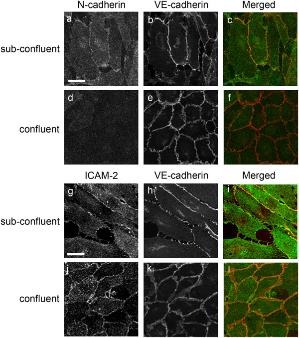
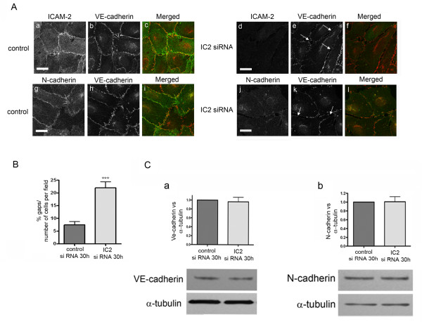
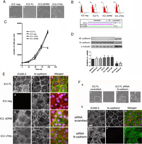
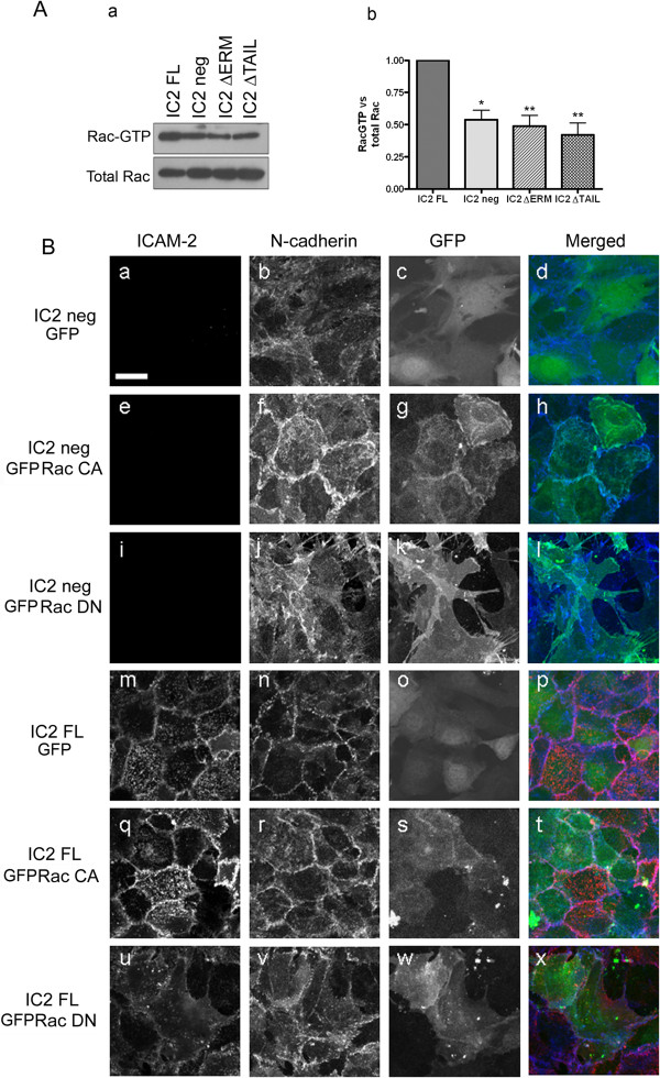
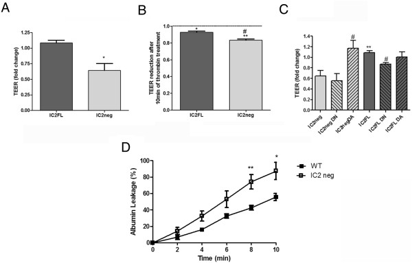
Results: In human umbilical vein endothelial cells (HUVEC), both VECad and NCad were found at nascent cell contacts of sub-confluent monolayers, but only VECad localized at the mature junctions of confluent monolayers. Inhibition of ICAM-2 expression by siRNA caused the appearance of small gaps at the junctions and a decrease in NCad junctional staining in sub-confluent monolayers. Endothelioma lines derived from WT or ICAM-2-deficient mice (IC2neg) lacked VECad and failed to form junctions, with loss of contact inhibition. Re-expression of full-length ICAM-2 (IC2 FL) in IC2neg cells restored contact inhibition through recruitment of NCad at the junctions. Mutant ICAM-2 lacking the binding site for ERM proteins (IC2 ΔERM) or the cytoplasmic tail (IC2 ΔTAIL) failed to restore junctions. ICAM-2-dependent Rac-1 activation was also decreased in these mutant cell lines. Barrier function, measured in vitro via transendothelial electrical resistance, was decreased in IC2neg cells, both in resting conditions and after thrombin stimulation. This was dependent on ICAM-2 signalling to the small GTPase Rac-1, since transendothelial electrical resistance of IC2neg cells was restored by constitutively active Rac-1. In vivo, thrombin-induced extravasation of FITC-labeled albumin measured by intravital fluorescence microscopy in the mouse cremaster muscle showed that permeability was increased in ICAM-2-deficient mice compared to controls.

Conclusions: These results indicate that ICAM-2 regulates endothelial barrier function and permeability through a pathway involving N-Cadherin, ERMs and Rac-1.


Figures

Figure 1
Figure 1
Distribution of ICAM-2, VECad and NCad in sub-confluent vs confluent HUVEC monolayers. Cells were seeded at low confluence (10000 cells/cm2) and cultured for 24 and 96 hours to achieve sub confluent and confluent endothelial monolayer respectively. For NCad/VECad co-staining (a-f), NCad was stained using mAb Cl32 anti-NCad followed by anti-mouse AlexaFluor488 (Green) and VECad was stained using mAb Cl55-7H1 anti-human VECad prelabelled with the Zenon® mouse IgG1 555 kit (Red). For ICAM-2/VECad co-staining (g-l), ICAM-2 was visualized using mAb BT-1 followed by anti-mouse AlexaFluor 488 (Green) and VECad was stained as described above. Co-staining for ICAM-2 and NCad was attempted with several antibodies and methods, however due to species incompatibility and the well known limitations of NCad antibodies available, satisfactory images could not be achieved. Bar = 25 μm.
Figure 2
Figure 2
Distribution of NCad in early endothelial cell-cell junctions is regulated by ICAM-2. A- Distribution of ICAM-2, VECad and NCad in HUVEC ~30 hours post siRNA treatment (a-i: control siRNA; d-l: ICAM-2 siRNA). Arrows indicate the gaps between cells that transiently appeared following ICAM-2 inhibition by siRNA. For ICAM-2/VECad co-staining (a-f), see details in Figure 1 legend. Bar = 25 μm B- Quantification of the number of gaps. The gaps were counted manually from 15 fields taken from confocal immunofluorescence images (233 μm x 233 μm, see panel B) in two independent experiments. Results are shown as % of gaps per number of cells per field. Error bars indicate mean ± S.E.M., n = 30. Statistical analysis (t-test: ***p < 0.001). C- VECad and NCad levels are unchanged 30 h post-treatment with ICAM2 siRNA. Quantification of VECad and NCad: Western blot quantification was performed by densitometry, normalized to α-tubulin. Error bars indicate mean ± s.e.m., n = 5.
Figure 3
Figure 3
ICAM-2 regulate N-Cad localization at cell-cell junction. A- Generation of mouse cardiac EC (MCEC) cell lines. Phase contrast image of IC2 neg (a), IC2FL (b), IC2 ΔERM (c) and IC2 ΔTAIL (d) cells. Bar = 150 μm. B- Analysis of ICAM-2 surface protein expression by FACS in IC2 neg (a), IC2 FL (b), IC2 ΔERM (c), IC2 ΔTAIL (d) cells; below, diagram showing the WT and mutant sequences of the constructs. FACS staining was performed using mAb 3C4 anti-mouse ICAM-2. C- Growth curves of MCEC cell lines. IC2 neg, IC2 ΔERM and IC2 ΔTAIL cells show loss of contact inhibition of cell growth. Results are represented as number of cells/cm2 over time (hours). D- Western blot analysis of NCad and VECad levels in IC2 neg, IC2 FL, IC2 ΔERM and IC2 ΔTAIL endothelioma lines and heart tissue from WT and IC2 deficient mice (ko). VECad and NCad were detected using mAb BV13 and mAb Cl32, respectively. Error bars indicate mean ± s.e.m., n = 3. E- Analysis of ICAM-2 and NCad distribution in IC2 FL (a-c), IC2 neg (d-f), IC2 ΔERM (g-i) and IC2 ΔTAIL (j-l) cells. ICAM-2 was stained with mAb 3C4 anti-mouse ICAM-2 followed by anti-rat AlexaFluor488 (Green). NCad was stained with mAb Cl32 anti-NCad followed by anti-mouse AlexaFluor555 (Red). Nuclei were stained using TOPRO-3 (Purple). Bar = 25 μm. F- Inhibition of NCad expression in IC2 FL cell line by siRNA resulted in disruption of cell-cell contacts and altered cell morphology. (a)- Phase contrast image of scrambled siRNA (a) and NCad siRNA (b) 48 hours post-transfection, Bar = 150 μm. (b)- Localization of ICAM-2 and NCad in IC2 FL cells treated with 48 hours scrambled (a-c) or NCad siRNA (d-f).
Figure 4
Figure 4
ERMs and Rac1 activity are involved in ICAM-2-dependent NCad recruitment and cell-cell contact. A- Rac1 activity in MCEC cell lines, as measured by the GST-PAK pull-down assay. (a) Representative Western blot analysis of Rac1 activity. Rac1-GTP pull-down with GST-PAK as well as total Rac1 were detected using mAb Cl23A8. (b) Quantification of Rac1 activity (Rac1-GTP/Total Rac1) in IC2 neg, IC2 FL, IC2 ΔERM, IC2 ΔTAIL cell lines. Error bars indicate mean ± s.e.m., n = 6. Statistical analysis (t-test: *p < 0.05, **p < 0.005). B- Effect of DN and CA Rac1 on the morphology and the distribution of NCad in the IC2 neg and IC2 FL lines. IC2 neg or IC2 FL were transfected with pEGFP control (a-d and m-p), pEGFP-V12 Rac1 (e-h and q-t) or pEGFP-V12-N17 Rac1 (i-l and u-x). Cells were co-stained for ICAM-2 and NCad. ICAM-2 was stained using the mAb 3C4 anti-mouse ICAM-2 followed by anti-rat AlexaFluor488 (pseudo colored Red). NCad was stained using mAb Cl32 anti-NCad followed by anti-mouse AlexaFluor555 (pseudo colored Blue). Bar = 25 μm.
Figure 5
Figure 5
In vitro and in vivo cell permeability assays. A- Cells were grown in full medium on gold electrodes until stable impedance was reached. TEER changes were recorded after 48 h and changes in TEER at baseline or B- after 10 min thrombin stimulation. Shown are average values -/+ s.e.m. of 4 independent experiments. C- Cells were grown in full medium on gold electrodes until stable impedance was reached. Cells were then infected with recombinant adenovirus encoding constitutively active (DA) or dominant negative (DN) myc-Rac1 at an m.o.i. of 400. After 4 h the virus was removed and cells starved to assess barrier development. TEER changes were recorded after 48 h. Shown are average values -/+ s.e.m of 4 independent experiments. D- In vivo permeability assay: albumin leakage in postcapillary venules from the cremaster muscles of WT and ICAM-2 KO (IC2 neg) mice in response to thrombin (2U/mL) during 10 min. Data are mean ± s.e.m. of n = 6 mice per group. t-test WT vs IC2-/- *p < 0.05, **p < 0.01.

References

    1. Bazzoni G, Dejana E. Endothelial cell-to-cell junctions: molecular organization and role in vascular homeostasis. Physiol Rev. 2004;84:869–901. doi: 10.1152/physrev.00035.2003. - DOI - PubMed
    1. Dejana E. Endothelial cell-cell junctions: happy together. Nat Rev Mol Cell Biol. 2004;5:261–270. doi: 10.1038/nrm1357. - DOI - PubMed
    1. Dejana E, Giampietro C. Vascular endothelial-cadherin and vascular stability. Curr Opin Hematol. 2012;19:218–223. doi: 10.1097/MOH.0b013e3283523e1c. - DOI - PubMed
    1. Kirkpatrick C, Peifer M. Not just glue: cell-cell junctions as cellular signaling centers. Curr Opin Genet Dev. 1995;5:56–65. doi: 10.1016/S0959-437X(95)90054-3. - DOI - PubMed
    1. Cavallaro U, Liebner S, Dejana E. Endothelial cadherins and tumor angiogenesis. Exp Cell Res. 2006;312:659–667. doi: 10.1016/j.yexcr.2005.09.019. - DOI - PubMed

Publication types

MeSH terms