

Cervical spine involvement in patients with juvenile idiopathic arthritis - MRI follow-up study
- PMID: 24593886
- PMCID: PMC3975869
- DOI: 10.1186/1546-0096-12-9
Cervical spine involvement in patients with juvenile idiopathic arthritis - MRI follow-up study
Abstract
Background: To describe MRI and clinical findings in patients with juvenile idiopathic arthritis with cervical spine involvement at onset and follow-up under therapy.
Methods: 13 patients with signs of cervical spine involvement in juvenile idiopathic arthritis with a median disease duration of 1.7 years were included in the study. Clinical records and MR images were retrospectively analyzed according to symptoms and findings concerning the cervical spine.
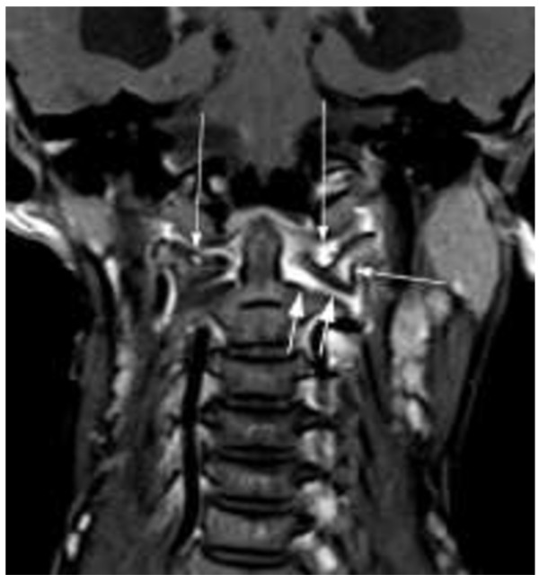
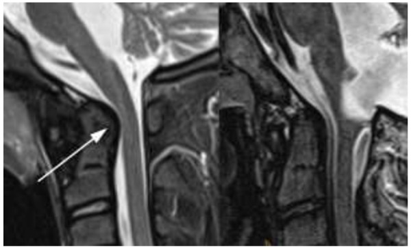
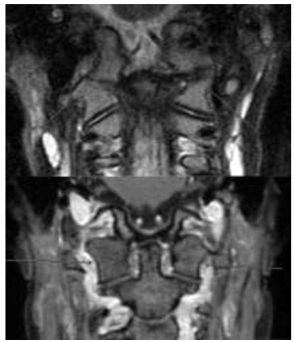
Results: At the onset of cervical spine involvement all patients showed limited range of motion, whereas only 5 of them complained of pain. In MR images joint hyperintensity, contrast enhancement, malalignment, ankylosis, erosion and narrowing of the spinal canal at cranio-cervical junction were found at 28, 32, 15, 2, 2 and 3 sites in 12 (93%), 13 (100%), 8 (62%), 2 (15%), 2 and 3 (20%) patients respectively. 3 of the 5 patients with pain (60%) showed ankylosis, erosions or narrowing of the spinal canal at cranio-cervical junction on MRI. At follow-up - after a median disease duration of cervical spine arthritis of 2.1 years and a variable duration of treatment with methotrexate (all patients) and biological agents (12 patients) - joint hyperintensity, enhancement and malalignment decreased to 15, 19 and 6 sites in 10 (77%), 11 (85%) and 3 (20%) patients respectively whereas ankylosis, erosion and narrowing of the spinal canal at cranio-cervical junction increased to 7, 6 and 4 sites in 3 (20%), 4 (31%) and 4 patients respectively. Pain was no longer reported, but 9 of 13 (69%) patients still had a limited range of motion with 6 of them (46%) showing skeletal changes on MRI.
Conclusions: This first MRI based follow-up study shows that cervical spine arthritis can follow a severe disease course in juvenile arthritis. While malalignments and inflammation sites decreased osseous changes with erosions, ankylosis, and narrowing of the spinal canal increased under treatment despite only minor subjective complaints. Therefore close MRI monitoring of these patients appears to be reasonable.
Figures
Similar articles
-
Clinical and MRI outcome of cervical spine lesions in children with juvenile idiopathic arthritis treated with anti-TNFα drugs early in disease course.Pediatr Rheumatol Online J. 2017 May 15;15(1):38. doi: 10.1186/s12969-017-0173-1. Pediatr Rheumatol Online J. 2017. PMID: 28506237 Free PMC article.
-
Juvenile rheumatoid arthritis: cervical spine involvement and MRI in early diagnosis.Turk J Pediatr. 1996 Apr-Jun;38(2):189-94. Turk J Pediatr. 1996. PMID: 8701483
-
The cervical spine in juvenile chronic arthritis.Spine J. 2002 Mar-Apr;2(2):89-94. doi: 10.1016/s1529-9430(02)00151-1. Spine J. 2002. PMID: 14588266
-
Cervical spine involvement early in the course of rheumatoid arthritis.Semin Arthritis Rheum. 2014 Jun;43(6):738-44. doi: 10.1016/j.semarthrit.2013.12.001. Epub 2013 Dec 12. Semin Arthritis Rheum. 2014. PMID: 24444595 Review.
-
Juvenile idiopathic arthritis overview and involvement of the temporomandibular joint: prevalence, systemic therapy.Oral Maxillofac Surg Clin North Am. 2015 Feb;27(1):1-10. doi: 10.1016/j.coms.2014.09.001. Oral Maxillofac Surg Clin North Am. 2015. PMID: 25483440 Review.
Cited by
-
Neuropsychological functioning and academic abilities in patients with juvenile idiopathic arthritis.Pediatr Rheumatol Online J. 2021 Apr 14;19(1):53. doi: 10.1186/s12969-021-00541-1. Pediatr Rheumatol Online J. 2021. PMID: 33853628 Free PMC article.
-
Advances in Musculoskeletal Imaging in Juvenile Idiopathic Arthritis.Biomedicines. 2022 Sep 27;10(10):2417. doi: 10.3390/biomedicines10102417. Biomedicines. 2022. PMID: 36289680 Free PMC article. Review.
-
Juvenile idiopathic arthritis - the role of imaging from a rheumatologist's perspective.Pediatr Radiol. 2018 Jun;48(6):785-791. doi: 10.1007/s00247-017-4014-7. Epub 2018 May 8. Pediatr Radiol. 2018. PMID: 29766250 Review.
-
Atraumatic atlantoaxial subluxation in pediatric enthesitis-related juvenile idiopathic arthritis: illustrative case.J Neurosurg Case Lessons. 2025 May 19;9(20):CASE25121. doi: 10.3171/CASE25121. Print 2025 May 19. J Neurosurg Case Lessons. 2025. PMID: 40388885 Free PMC article.
-
A clinical perspective on imaging in juvenile idiopathic arthritis.Pediatr Radiol. 2024 Apr;54(4):490-504. doi: 10.1007/s00247-023-05815-2. Epub 2023 Nov 28. Pediatr Radiol. 2024. PMID: 38015293 Free PMC article. Review.
References
-
- Neva MH, Kaarela K, Kauppi M. Prevalence of radiological changes in the cervical spine–a cross sectional study after 20 years from presentation of rheumatoid arthritis. J Rheumatol. 2000;27(1):90–93. - PubMed
-
- Hensinger RN, DeVito PD, Ragsdale CG. Changes in the cervical spine in juvenile rheumatoid arthritis. J Bone Joint Surg Am. 1986;86(2):189–198. - PubMed
Publication types
MeSH terms
Substances
LinkOut - more resources
Full Text Sources
Other Literature Sources
Medical
