. 2014 Apr;21(4):472-80.
doi: 10.1016/j.acra.2013.12.012.

Altered median nerve deformation and transverse displacement during wrist movement in patients with carpal tunnel syndrome

Affiliations

Altered median nerve deformation and transverse displacement during wrist movement in patients with carpal tunnel syndrome

Yuexiang Wang et al. Acad Radiol. 2014 Apr.

Abstract

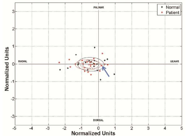
Rationale and objectives: Carpal tunnel syndrome (CTS) is the most common peripheral nerve entrapment syndrome. Strong pinch or grip with wrist flexion has been considered a risk factor for CTS. Studying median nerve displacement during wrist movements may provide useful information about median nerve kinematic changes in patients with CTS. The purpose of this study was to evaluate the deformability and mobility of the median nerve in patients with CTS compared to healthy subjects.

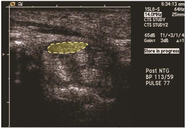
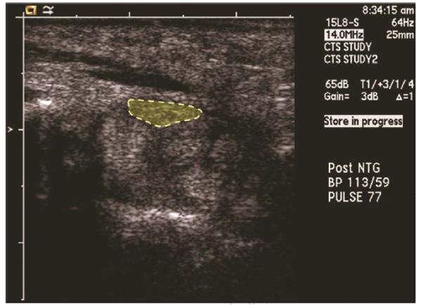
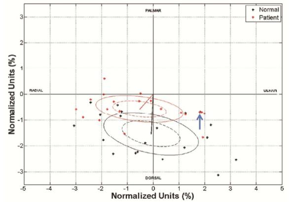
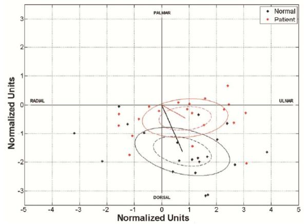
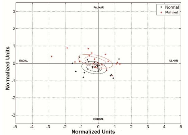
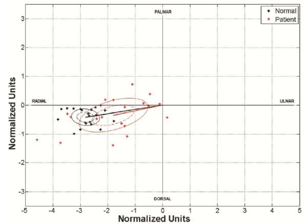
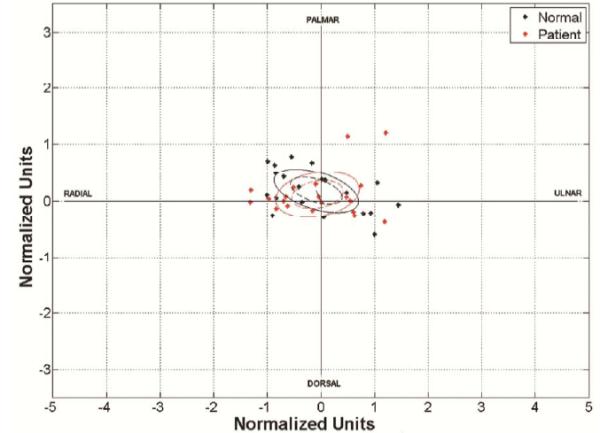
Materials and methods: Dynamic ultrasound images were obtained in 20 affected wrists of 13 patients with CTS. Results were compared to complementary data obtained from both wrists of 10 healthy subjects reported in a previous study. Shape and position of initial and final median nerve were measured and analyzed for six defined wrist movements. The deformation ratios for each movement were defined as the median nerve area, perimeter, and circularity of the final position normalized by respective values assessed in the initial position. The median nerve displacement vector and magnitude were also calculated.

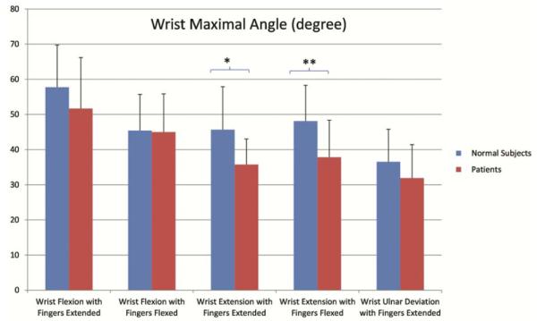
Results: The deformation ratio for circularity was significantly less in patients with CTS compared to healthy subjects during wrist flexion (P < .05). The mean vector of median nerve displacement during wrist flexion was significantly different between patients with CTS and healthy subjects (P < .05). The displacement magnitude of the median nerve was found to be less in patients with CTS compared to healthy subjects during most movements, with the exception of wrist extension with fingers extended.

Conclusions: Patients with CTS differ from normal subjects with regard to mobility and deformability of the median nerve.

Keywords: Ultrasound; carpal tunnel syndrome; median nerve.


Figures

Figure 1
Figure 1
The comparison of the wrist angles between CTS patients and healthy subjects. The maximal wrist angle during wrist extension with fingers extended or flexed in CTS patients was significantly lower than those in healthy subjects.
Figure 2
Figure 2
The median nerve at the carpal tunnel in a patient with CTS. A. At the start of wrist maximal flexion with fingers extended, the enlarged median nerve (dashed ellipse) was located between the flexor retinaculum and flexor tendons on transverse ultrasound. B. At the end of the movement the median nerve (dashed ellipse) was still located between the flexor retinaculum and flexor tendons and had moved only slightly towards the ulnar side of the carpal tunnel.
Figure 2
Figure 2
The median nerve at the carpal tunnel in a patient with CTS. A. At the start of wrist maximal flexion with fingers extended, the enlarged median nerve (dashed ellipse) was located between the flexor retinaculum and flexor tendons on transverse ultrasound. B. At the end of the movement the median nerve (dashed ellipse) was still located between the flexor retinaculum and flexor tendons and had moved only slightly towards the ulnar side of the carpal tunnel.
Figure 3
Figure 3
The median nerve displacement vectors in CTS patients. Solid ellipses represent standard deviation, dashed ellipses represent 95% confidence limits and the radial line represents the mean vector for each group. A. Wrist flexion with fingers extended. The arrow indicates two superimposed points from a patient and a healthy subject respectively. B. Wrist flexion with fingers flexed. C. Wrist extension with fingers extended. D. Wrist ulnar deviation with fingers extended. E. Finger flexion. F. Wrist extension with fingers flexed. The arrow indicates two superimposed points from a patient and a healthy subject respectively.
Figure 3
Figure 3
The median nerve displacement vectors in CTS patients. Solid ellipses represent standard deviation, dashed ellipses represent 95% confidence limits and the radial line represents the mean vector for each group. A. Wrist flexion with fingers extended. The arrow indicates two superimposed points from a patient and a healthy subject respectively. B. Wrist flexion with fingers flexed. C. Wrist extension with fingers extended. D. Wrist ulnar deviation with fingers extended. E. Finger flexion. F. Wrist extension with fingers flexed. The arrow indicates two superimposed points from a patient and a healthy subject respectively.

References

    1. Nakamichi K, Tachibana S. Restricted motion of the median nerve in carpal tunnel syndrome. J Hand Surg Br. 1995;20(4):460–4. - PubMed
    1. Ikeda K, Sarnura N, Tornita K. Segmental carpal canal pressure in patients with carpal tunnel syndrome. J Hand Surg Am. 2006;31A(6):925–9. - PubMed
    1. Werner CO, Elmqvist D, Ohlin P. Pressure and nerve lesion in the carpal tunnel. Acta Orthop Scand. 1983;54(2):312–6. - PubMed
    1. Coppieters MW, Alshami AM. Longitudinal excursion and strain in the median nerve during novel nerve gliding exercises for carpal tunnel syndrome. J Orthop Res. 2007;25(7):972–80. - PubMed
    1. Hough AD, Moore AP, Jones MP. Reduced longitudinal excursion of the median nerve in carpal tunnel syndrome. Arch Phys Med Rehabil. 2007;88(5):569–76. - PubMed

Publication types