. 2014 Mar 4;9(3):e89894.
doi: 10.1371/journal.pone.0089894. eCollection 2014.

Effect of low level laser therapy on chronic compression of the dorsal root ganglion

Affiliations

Effect of low level laser therapy on chronic compression of the dorsal root ganglion

Yi-Jen Chen et al. PLoS One. .

Abstract

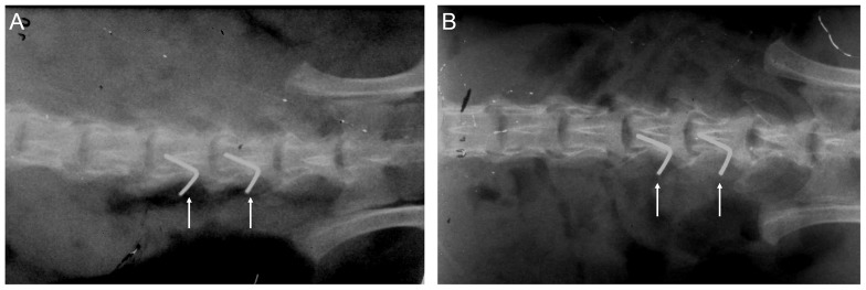
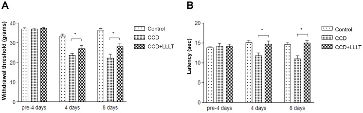
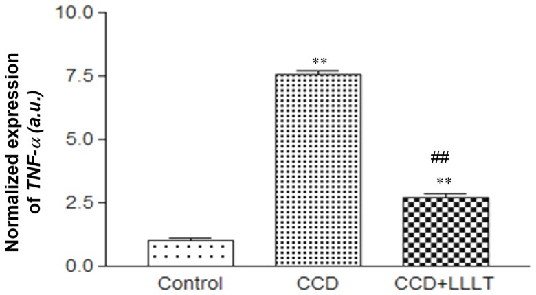
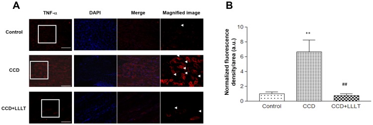
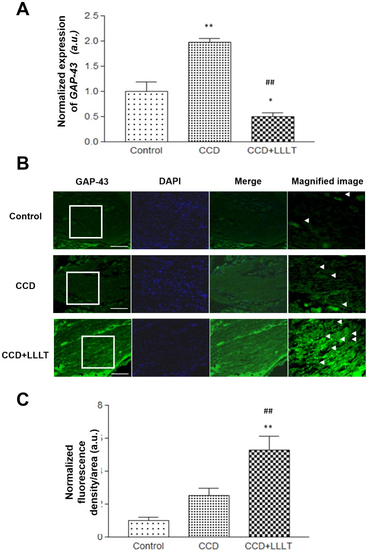
Dorsal root ganglia (DRG) are vulnerable to physical injury of the intervertebral foramen, and chronic compression of the DRG (CCD) an result in nerve root damage with persistent morbidity. The purpose of this study was to evaluate the effects of low level laser therapy (LLLT) on the DRG in a CCD model and to determine the mechanisms underlying these effects. CCD rats had L-shaped stainless-steel rods inserted into the fourth and fifth lumbar intervertebral foramen, and the rats were then subjected to 0 or 8 J/cm2 LLLT for 8 consecutive days following CCD surgery. Pain and heat stimuli were applied to test for hyperalgesia following CCD. The levels of TNF-α, IL-1β and growth-associated protein-43 (GAP-43) messenger RNA (mRNA) expression were measured via real-time PCR, and protein expression levels were analyzed through immunohistochemical analyses. Our data indicate that LLLT significantly decreased the tolerable sensitivity to pain and heat stimuli in the CCD groups. The expression levels of the pro-inflammatory cytokines TNF-α and IL-1β were increased following CCD, and we found that these increases could be reduced by the application of LLLT. Furthermore, the expression of GAP-43 was enhanced by LLLT. In conclusion, LLLT was able to enhance neural regeneration in rats following CCD and improve rat ambulatory behavior. The therapeutic effects of LLLT on the DRG during CCD may be exerted through suppression of the inflammatory response and induction of neuronal repair genes. These results suggest potential clinical applications for LLLT in the treatment of compression-induced neuronal disorders.


Conflict of interest statement

Competing Interests: The authors have declared that no competing interests exist.

Figures

Figure 1
Figure 1. A soft X-ray radiographic analysis of CCD, both immediately following CCD surgery (A) and 8 days post-surgery (B); the arrows indicate the position of the L-shaped needles.
Figure 2
Figure 2. LLLT reduced CCD-mediated mechanical and thermal hyperalgesia.
The behavioral patterns of rats in the control, CCD and CCD+LLLT groups were monitored at the indicated times (n = 6). (A) Foot withdrawal threshold responses to mechanical stimuli. (B) Foot withdrawal latencies in response to thermal stimuli. The levels of statistical significance are as follows: *, p<0.05.
Figure 3
Figure 3. LLLT suppressed the mRNA expression level of CCD-induced TNF-α.
The level of TNF-α mRNA expression was analyzed using the 2−ΔCT method and normalized to the Control group. The levels of statistical significance are as follows: **, p<0.01 relative to the Control group; ##, p<0.01 relative to the CCD group.
Figure 4
Figure 4. LLLT suppressed the protein expression levels of CCD-induced inflammatory cytokines.
(A) Fluorescence images of TNF-α staining from the Control and CCD groups with or without LLLT were analyzed. Representative images from the Control (upper row), CCD (middle row) and CCD+LLLT (bottom row) groups are shown. TNF-α signals are shown in the left column (red). Images of nuclear staining with DAPI are presented in the middle-left column (blue), and merged fluorescence images are shown in the middle-right column. Magnified image of TNF-α are provided in the right column (magnification 400×) and representative signals are indicated by arrowheads. Scale bars, 10 µm. (B) A graph displaying the quantification of TNF-α fluorescence. The levels of statistical significance are as follows: **, p<0.01 relative to the Control group; ##, p<0.01 relative to the CCD group.
Figure 5
Figure 5. LLLT increased GAP-43 expression in a CCD model.
(A) The levels of GAP-43 mRNA expression were analyzed using the 2−ΔCT method and normalized to the Control group. (B) Fluorescence images of GAP-43 staining from the Control or CCD groups with or without LLLT were analyzed. Representative images from the Control (upper row), CCD (middle row) and CCD+LLLT (bottom row) groups are shown. GAP-43 signals are presented in the left column (green). Images of nuclear staining with DAPI are provided in the middle-left column (blue), and merged fluorescence images are shown in the middle-right column Magnified image of GAP-43 are provided in the right column (magnification 400×) and representative signals are indicated by arrowheads. Scale bars, 10 µm. (C) A graph displaying the quantification of GAP-43 fluorescence. The levels of statistical significance are as follows: *, p<0.05 and **, p<0.01 relative to the control group; #, p<0.05 and ##, p<0.01 relative to the CCD groups.

References

    1. Rydevik BL, Myers RR, Powell HC (1989) Pressure increase in the dorsal root ganglion following mechanical compression. Closed compartment syndrome in nerve roots. Spine (Phila Pa 1976) 14: 574–576. - PubMed
    1. Gilchrist RV, Slipman CW, Bhagia SM (2002) Anatomy of the intervertebral foramen. Pain Physician 5: 372–378. - PubMed
    1. Schaeffer V, Meyer L, Patte-Mensah C, Mensah-Nyagan AG (2010) Progress in dorsal root ganglion neurosteroidogenic activity: basic evidence and pathophysiological correlation. Prog Neurobiol 92: 33–41. - PubMed
    1. de Souza Grava AL, Ferrari LF, Defino HL (2012) Cytokine inhibition and time-related influence of inflammatory stimuli on the hyperalgesia induced by the nucleus pulposus. Eur Spine J 21: 537–545. - PMC - PubMed
    1. Ozaktay AC, Cavanaugh JM, Asik I, DeLeo JA, Weinstein JN (2002) Dorsal root sensitivity to interleukin-1 beta, interleukin-6 and tumor necrosis factor in rats. Eur Spine J 11: 467–475. - PMC - PubMed

Publication types

MeSH terms