. 2014 Mar 3;9(3):e90389.
doi: 10.1371/journal.pone.0090389. eCollection 2014.

Endoplasmic reticulum protein ERp46 in renal cell carcinoma

Affiliations

Endoplasmic reticulum protein ERp46 in renal cell carcinoma

Wilhelmina C M Duivenvoorden et al. PLoS One. .

Abstract

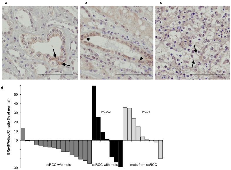
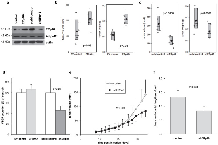
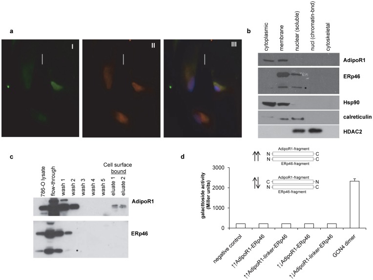
An established inverse clinical correlation between serum adiponectin levels and renal cell carcinoma (RCC) aggressiveness exists. We have recently demonstrated that adiponectin suppresses clear cell RCC (ccRCC) progression through interaction with its receptor, adiponectin receptor 1 (AdipoR1). ERp46 has been shown to inhibit adiponectin signaling via interaction with AdipoR1 in HeLa cells. However, the expression of ERp46 in RCC has not been described thus far. The objectives of this study were to investigate ERp46 in RCC, its expression, its effects on RCC growth in a mouse model and whether it interacts with AdipoR1. We demonstrated a higher ERp46/AdipoR1 expression ratio in metastatic compared to non-metastatic ccRCC, as determined by immunohistochemistry of tissue microarrays and subsequent image analysis. When ERp46 was stably knocked down using shRNA or overexpressed in murine RCC RAG cells, RCC growth after subcutaneous injection in BALB/c nude mice was inhibited and accelerated, respectively. In vitro analysis to determine the molecular interaction between AdipoR1 and ERp46 included co-immunoprecipitation using human ccRCC 786-O cells and a bacterial adenylate cyclase-based two hybrid system and demonstrated no sustained AdipoR1-ERp46 interaction. This is the first report to suggest a role for ERp46 as a potential therapeutic target in RCC given its expression profile in human RCC samples and its effect on in vivo RCC growth. Since a stable interaction with AdipoR1 could not be established, we suggest that the tumorigenic properties of ERp46 in RCC cells are not related to an inhibitory modulation of AdipoR1.


Conflict of interest statement

Competing Interests: The authors have declared that no competing interests exist.

Figures

Figure 1
Figure 1. ERp46 expression is increased in metastatic human ccRCC tissue.
ERp46-immunohistochemistry of human normal kidney tissue demonstrates (a) granular cytoplasmic staining typical for ER (examples indicated by the arrows), but also (b) nuclear staining (arrow heads). (c) ERp46-staining of human ccRCC showing plasma membrane staining (arrows). Original magnification 630x; Bar = 100 µm. (d) The ratio of ERp46/AdipoR1 protein expression in specimens of ccRCC patients was significantly increased in primary ccRCC from patients with metastasis (p = 0.002) and in metastatic tissue (p = 0.04). ERp46 and AdipoR1 protein expression was quantified by image analysis (H-score). The AdipoR1 and ERp46 protein expression in the cancer was normalized to their expression in normal tissue. The ratios obtained from the patients with primary ccRCC without distant metastasis are represented by the dark grey bars, the black ones represent the primary ccRCC samples from patients with metastasis, the light grey ones are from ccRCC metastatic samples.
Figure 2
Figure 2. ERp46 supports in vivo tumor growth.
(a) ERp46-manipulated subclones of mouse RCC RAG cells demonstrated a 4-fold increase in ERp46 protein expression in ERp46-overexpressing (ERp46+) cells, and a 80% knockdown of ERp46 protein expression in cells stably expressing shRNA specific for ERp46 (shERp46) compared to corresponding control transfected cells. There was no difference in AdipoR1 expression. ERp46 and AdipoR1 were detected by Western blot analysis. The protein expression of β-actin served as loading control. (b) In vivo growth of different ERp46-manipulated stable subclones of mouse RCC RAG cells. Tumor volume and weight from mice (n = 7/group) injected with ERp46+-RAG cells is significantly higher than from mice injected with empty vector (EV) control-transfected cells (p = 0.02 and 0.03, respectively). Individual values (○) and mean (♦) are shown, the box indicates the 95% confidence interval. (c) Tumor volume and weight from mice (n = 7/group) subcutaneously injected with shERp46-RAG cells is significantly lower than from mice injected with cells stably transfected with scrambled control shRNA (p = 0.0006 and 0.0001, respectively). Individual values (○) and mean (♦) are shown, the box indicates the 95% confidence interval. (d) Serum VEGF of the mice subcutaneously injected with ERp46-manipulated RAG cells 35 days after injection (n = 7/group). Serum VEGF is significantly lower in shERp46 RAG cell-injected mice compared to corresponding control mice (p = 0.02). Data represent mean ±95% confidence intervals. (e) Longitudinal tumor growth of mouse RCC RAG cells in vivo. Mice were subcutaneously injected with 2×10⁶ RAG cells and treated systemically (intraperitoneally) every second day with shRNA specific for ERp46 (shERp46) or scrambled control shRNA (control) (n = 10/group) using the in-vivojetPEI delivery agent (p = 0.001; ANOVA). Data represent mean ±95% confidence intervals. (f) At sacrifice (35 days), the linear endothelial length as determined in CD31-stained subcutaneous tumors is significantly lower (p = 0.003) in shERp46-treated RCC RAG cell-injected mice (n = 10) compared to mice treated with shControl (n = 10). Data represent mean ±95% confidence intervals.
Figure 3
Figure 3. Co-localization of ERp46 and AdipoR1 in human ccRCC 786-O cells, but no interaction.
(a) Immunocytochemical staining for ERp46 (I, green), or AdipoR1 (II, red). The merged image (III) demonstrates yellow signal which indicates co-localization. Cells were counterstained with DAPI (blue). (b) Subcellular protein fractionation. Equal portions of each fractionated cellular extract were analyzed by Western blot using specific antibodies against AdipoR1 and ERp46. Antibodies directed against Hsp90 (cytoplasmic), calreticulin (plasma membrane) and HDAC2 (nuclear) served as fractionation controls. AdipoR1 is detected in the cytoplasmic and plasma membrane fractions, ERp46 in the nuclear soluble and membrane fraction. The asterisk indicates an ERp46 degradation product or possibly the shorter ERp46 isoform 3. (c). Western blot analyses for AdipoR1 and ERp46-specific extraction and isolation from 3×10⁶ 786-O cells. Presence of AdipoR1 in the elution fraction confirms that AdipoR1 is cell surface exposed. Absence of ERp46 in the elution fraction indicates most likely that it is either not cell surface exposed or not tightly bound to a cell surface protein. (d) Bacterial adenylate cyclase-based two-hybrid assay (BACTH) used to determine the interaction between ERp46 and AdipoR1. The N-termini of AdipoR1 and ERp46 were expressed as fusion to the T18 and T25 domains of the adenylate cyclase. Interaction was quantified via cAMP/CAP-induced β-galactosidase activity. The pUT18C strain producing AdipoR1N fused to T25 domain of adenylate cyclase served as negative control. Other controls (plasmid, ERp46N with linker, ERp46N fused to T18) were also negative. No interaction between the N-termini of ERp46 and AdipoR1 was observed. T18 and T25 fused to interacting leucine zippers from GCN4 served as positive control. Data are from three independent repetitions and error bars indicate standard deviation. Depending on the fusion direction of AdipoR1N and ERp46N to T18 and T25, a “parallel” and “anti-parallel” orientation of the N-termini is captured, shown schematically.

References

    1. Klinghoffer Z, Yang B, Kapoor A, Pinthus JH (2009) Obesity and renal cell carcinoma: epidemiology, underlying mechanisms and management considerations. Expert Rev Anticancer Ther 9: 975–987. - PubMed
    1. Diez JJ, Iglesias P (2003) The role of the novel adipocyte-derived hormone adiponectin in human disease. Eur J Endocrinol 148: 293–300. - PubMed
    1. Gavrila A, Chan JL, Yiannakouris N, Kontogianni M, Miller LC, et al. (2003) Serum adiponectin levels are inversely associated with overall and central fat distribution but are not directly regulated by acute fasting or leptin administration in humans: cross-sectional and interventional studies. J Clin Endocrinol Metab 88: 4823–4831. - PubMed
    1. Trujillo ME, Scherer PE (2005) Adiponectin—journey from an adipocyte secretory protein to biomarker of the metabolic syndrome. J Intern Med 257: 167–175. - PubMed
    1. Pinthus JH, Kleinmann N, Tisdale B, Chatterjee S, Lu JP, et al. (2008) Lower plasma adiponectin levels are associated with larger tumor size and metastasis in clear-cell carcinoma of the kidney. Eur Urol 54: 866–873. - PubMed

Publication types

Substances