The diagnostic accuracy of ultrasonography versus endoscopy for primary nasopharyngeal carcinoma
- PMID: 24594807
- PMCID: PMC3940890
- DOI: 10.1371/journal.pone.0090412
The diagnostic accuracy of ultrasonography versus endoscopy for primary nasopharyngeal carcinoma
Erratum in
-
The diagnostic accuracy of ultrasonography versus endoscopy for primary nasopharyngeal carcinoma.PLoS One. 2014 Jun 2;9(6):e99679. doi: 10.1371/journal.pone.0099679. eCollection 2014. PLoS One. 2014. PMID: 24887439 Free PMC article.
Abstract
Objective: To compare the accuracy of ultrasonography (US) with the current clinical standard of endoscopy for a diagnosis of nasopharyngeal carcinoma (NPC).
Methods: A total of 150 patients suspected of having NPC underwent US and endoscopy. A diagnosis was obtained from an endoscopic biopsy collected from each suspected tumor and was compared with a biopsy obtained from a normal nasopharynx. The diagnostic accuracy of US and endoscopy for NPC was evaluated using receiver operating curve (ROC) analysis performed by MedCalc Software.
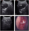
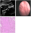
Results: The sensitivity, specificity, and accuracy of US versus endoscopy for this cohort were 90.1%, 84.8%, and 87.3% for US, and 88.7%, 97.5%, and 93.3% for endoscopy, respectively. Both US and endoscopy exhibited good diagnostic accuracy for NPC with area under the curve (AUC) values of 0.929 and 0.938, respectively. However, this difference was not significant (Z = 0.36, P = 0.72).
Conclusion: US is a useful tool for the detection of tumors in endoscopically suspicious nasopharynx tissues, and also for the detection of subclinical tumors in endoscopically normal nasopharynx tissues.
Conflict of interest statement
Figures


Similar articles
-
The diagnostic accuracy of ultrasonography versus endoscopy for primary nasopharyngeal carcinoma.PLoS One. 2014 Jun 2;9(6):e99679. doi: 10.1371/journal.pone.0099679. eCollection 2014. PLoS One. 2014. PMID: 24887439 Free PMC article.
-
Primary nasopharyngeal carcinoma: diagnostic accuracy of MR imaging versus that of endoscopy and endoscopic biopsy.Radiology. 2011 Feb;258(2):531-7. doi: 10.1148/radiol.10101241. Epub 2010 Dec 3. Radiology. 2011. PMID: 21131580
-
Detection of Nasopharyngeal Carcinoma by MR Imaging: Diagnostic Accuracy of MRI Compared with Endoscopy and Endoscopic Biopsy Based on Long-Term Follow-Up.AJNR Am J Neuroradiol. 2015 Dec;36(12):2380-5. doi: 10.3174/ajnr.A4456. Epub 2015 Aug 27. AJNR Am J Neuroradiol. 2015. PMID: 26316564 Free PMC article.
-
Diagnostic Value of Serum Epstein-Barr Virus Capsid Antigen-IgA for Nasopharyngeal Carcinoma: a Meta-Analysis Based on 21 Studies.Clin Lab. 2016;62(6):1155-66. doi: 10.7754/clin.lab.2015.151122. Clin Lab. 2016. PMID: 27468579 Review.
-
Diagnostic Capacity of RASSF1A Promoter Methylation as a Biomarker in Tissue, Brushing, and Blood Samples of Nasopharyngeal Carcinoma.EBioMedicine. 2017 Apr;18:32-40. doi: 10.1016/j.ebiom.2017.03.038. Epub 2017 Apr 2. EBioMedicine. 2017. PMID: 28396012 Free PMC article. Review.
Cited by
-
Prognostic value of inflammation-based prognostic scores on outcome in patients undergoing continuous ambulatory peritoneal dialysis.BMC Nephrol. 2018 Oct 26;19(1):297. doi: 10.1186/s12882-018-1092-1. BMC Nephrol. 2018. PMID: 30367618 Free PMC article.
-
Contrast-enhanced Ultrasound in evaluating of angiogenesis and tumor staging of nasopharyngeal carcinoma in nude mice.PLoS One. 2019 Aug 23;14(8):e0221638. doi: 10.1371/journal.pone.0221638. eCollection 2019. PLoS One. 2019. PMID: 31442259 Free PMC article.
-
The diagnostic accuracy of ultrasonography versus endoscopy for primary nasopharyngeal carcinoma.PLoS One. 2014 Jun 2;9(6):e99679. doi: 10.1371/journal.pone.0099679. eCollection 2014. PLoS One. 2014. PMID: 24887439 Free PMC article.
-
Pathologic Evaluation of Routine Nasopharynx Punch Biopsy in the Adult Population: Is It Really Necessary?Clin Exp Otorhinolaryngol. 2017 Sep;10(3):283-287. doi: 10.21053/ceo.2015.01256. Epub 2016 Jul 27. Clin Exp Otorhinolaryngol. 2017. PMID: 27459201 Free PMC article.
-
Clinical evaluation of vascular normalization induced by recombinant human endostatin in nasopharyngeal carcinoma via dynamic contrast-enhanced ultrasonography.Onco Targets Ther. 2018 Nov 8;11:7909-7917. doi: 10.2147/OTT.S181842. eCollection 2018. Onco Targets Ther. 2018. PMID: 30510431 Free PMC article.
References
-
- Chan AT, Felip E (2009) ESMO Guidelines Working Group (2009) Nasopharyngeal cancer: ESMO clinical recommendations for diagnosis, treatment and follow-up. Ann Oncol 20: 123–125. - PubMed
-
- King AD, Vlantis AC, Bhatia KS, Zee BC, Woo JK, et al. (2011) Primary nasopharyngeal carcinoma: diagnostic accuracy of MR imaging versus that of endoscopy and endoscopic biopsy. Radiology 258: 531–537. - PubMed
-
- Wei WI, Sham JS, Zong YS, Choy D, Ng MH (1991) The efficacy of fiberoptic endoscopic examination and biopsy in the detection of early nasopharyngeal carcinoma. Cancer 67: 3127–3130. - PubMed
-
- Abdel Khalek Abdel Razek A, King A (2012) MRI and CT of nasopharyngeal carcinoma. AJR Am J Roentgenol 198: 11–18. - PubMed
Publication types
MeSH terms
LinkOut - more resources
Full Text Sources
Other Literature Sources
Medical

