. 2014 Mar 3;9(3):e90727.
doi: 10.1371/journal.pone.0090727. eCollection 2014.

Osteosarcoma microenvironment: whole-slide imaging and optimized antigen detection overcome major limitations in immunohistochemical quantification

Affiliations

Osteosarcoma microenvironment: whole-slide imaging and optimized antigen detection overcome major limitations in immunohistochemical quantification

Pierre Kunz et al. PLoS One. .

Abstract

Background: In osteosarcoma survival rates could not be improved over the last 30 years. Novel biomarkers are warranted to allow risk stratification of patients for more individual treatment following initial diagnosis. Although previous studies of the tumor microenvironment have identified promising candidates, novel biomarkers have not been translated into routine histopathology. Substantial difficulties regarding immunohistochemical detection and quantification of antigens in decalcified and heterogeneous osteosarcoma might largely explain this translational short-coming. Furthermore, we hypothesized that conventional hot spot analysis is often not representative for the whole section when applied to heterogeneous tissues like osteosarcoma. We aimed to overcome these difficulties for major biomarkers of the immunovascular microenvironment.

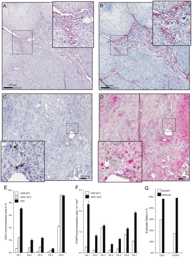
Methods: Immunohistochemistry was systematically optimized for cell surface (CD31, CD8) and intracellular antigens (FOXP3) including evaluation of 200 different antigen retrieval conditions. Distribution patterns of these antigens were analyzed in formalin-fixed and paraffin-embedded samples from 120 high-grade central osteosarcoma biopsies and computer-assisted whole-slide analysis was compared with conventional quantification methods including hot spot analysis.

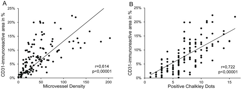
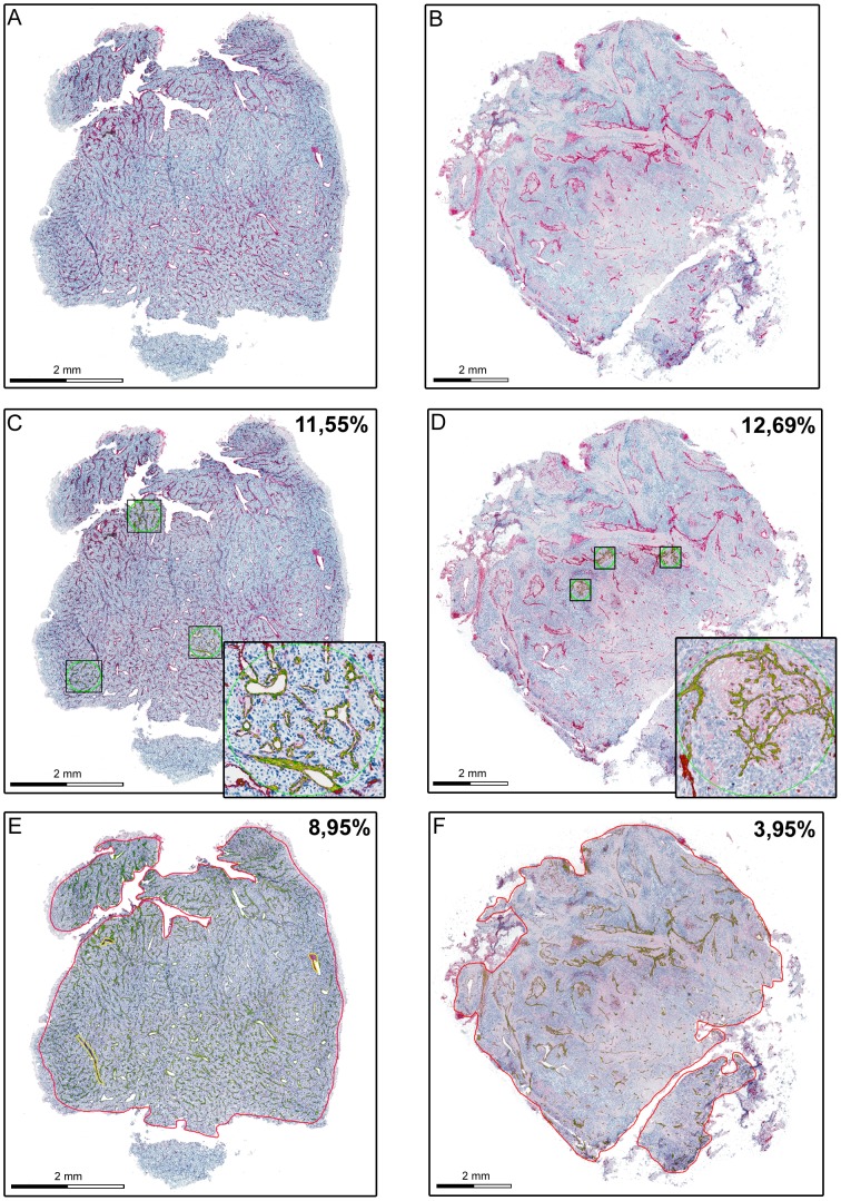
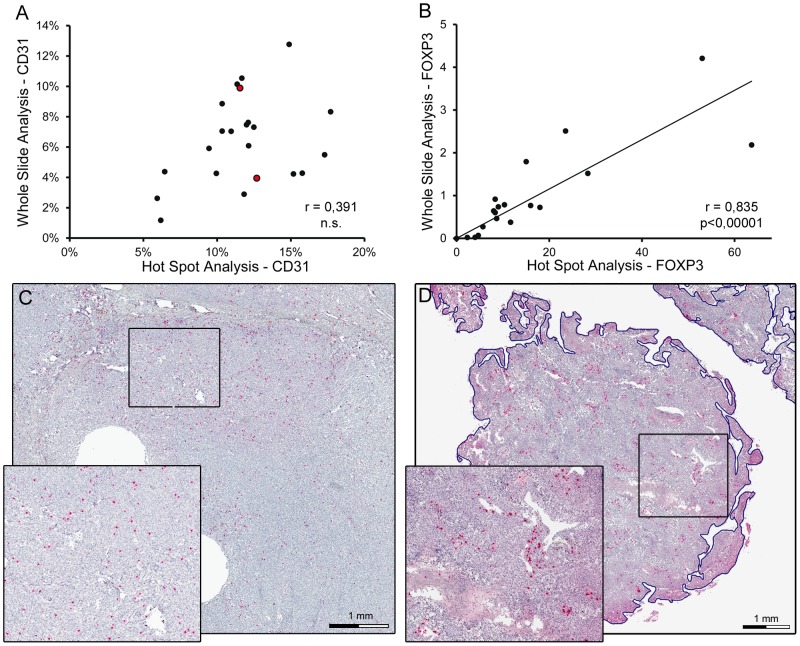
Results: More than 96% of osteosarcoma samples were positive for all antigens after optimization of immunohistochemistry. In contrast, standard immunohistochemistry retrieved false negative results in 35-65% of decalcified osteosarcoma specimens. Standard hot spot analysis was applicable for homogeneous distributed FOXP3+ and CD8+ cells. However, heterogeneous distribution of vascular CD31 did not allow reliable quantification with hot spot analysis in 85% of all samples. Computer-assisted whole-slide analysis of total CD31- immunoreactive area proved as the most appropriate quantification method.

Conclusion: Standard staining and quantification procedures are not applicable in decalcified formalin-fixed and paraffin-embedded samples for major parameters of the immunovascular microenvironment in osteosarcoma. Whole-slide imaging and optimized antigen retrieval overcome these limitations.


Conflict of interest statement

Competing Interests: The authors have declared that no competing interests exist.

Figures

Figure 1
Figure 1. Effects of different staining conditions on the quantification of immunovascular markers in osteosarcoma.
(A) Formalin-fixed and paraffin-embedded osteosarcoma sample after CD31 staining with standard heat induced epitope retrieval at 98°C and (B) with optimized enzymatic epitope retrieval. CD31- immunoreactive cells show red cell surface staining. Section was counterstained by hematoxylin. Insert shows 2-fold magnification of indicated area. (C) Formalin-fixed and paraffin-embedded osteosarcoma sample after FOXP3 staining with standard heat induced epitope retrieval at 98°C and (D) with optimized epitope retrieval at 127°C. FOXP3 immunoreactive cells (arrows) show red nuclear staining. Section was counterstained with hematoxylin. Insert shows 3.8 fold magnification of indicated area. (E) Percentage of CD31 immunoreactive area was assessed by computer-assisted whole-slide quantification after heat induced epitope retrieval (HIER) at 98°C, HIER at 127° and enzymatic epitope retrieval (EER) with Hyaluronidase and Pronase in five representative osteosarcoma samples (OS). Error bars indicate interobserver variability. (F) Only seven out of the 20 tested osteosarcoma samples showed FOXP3 immunoreactive cells after standard heat induced epitope retrieval at 98°C (not shown). Density of FOXP3-immunoreactive cells (numbers/0.1 mm2) was determined in these seven sections by whole-slide quantification after HIER at 98°C and for HIER at 127°C. Error bars indicates interobserver variability. OS = osteosarcoma sample. (G) Percentage of evaluable slides after standard and optimized immunohistochemical staining for CD31 and FOXP3.
Figure 2
Figure 2. Correlation of common vessel quantification methods in osteosarcoma.
Correlation of vessel quantification derived by total CD31-immunoreactive area and micro vessel density (A), respectively total CD31- immunoreactive area and Chalkley count (B) within 120 predefined spots with 0.26 mm2 area/spot in 20 representative osteosarcoma sample; r indicates Pearson correlation coefficient.
Figure 3
Figure 3. Effect of heterogeneous vessel distribution on vessel quantification in osteosarcoma.
Representative whole-slide scans of formalin-fixed and paraffin-embedded osteosarcoma samples with homogeneously scattered (A,C,E) and hot spot distributed intratumor vascularization (B,D,F) Quantification of tumor vascularization was either performed by hot spot analysis within three circular hot spots with 0.26 mm2 area/hot spot (C and D) or whole-slide analysis of CD31-immunoreactive area (E and F). CD31 immunoreactivity is shown in red. By digital image analysis detected CD31-immunoreactive area is annotated in green (automated mark-up image). Indicated values represent the percentage of immunoreactive area within the analyzed regions (three hot spots in C and D, whole slide in E and F). Inserts show a 5-fold (C), respectively 8-fold (D) magnification of indicated regions. Sections were counterstained by hematoxylin.
Figure 4
Figure 4. Correlation of hot spot analysis and whole slide analysis of immunovascular markers in osteosarcoma.
(A) Correlation between hot spot and whole slide analyses of CD31-immunoreactive area (in %) of 20 representative osteosarcoma samples. Circles filled in red represent the two specimens shown in Figure 3. Pearson correlation coefficient is indicated by r, n.s. =  not significant. (B) Correlation between hot spot and whole slide analyses of FOXP3 cell density (cells per 0.1 mm2) of 20 representative osteosarcoma samples. Pearson correlation coefficient is indicated by r and significance by p. (C) Representative osteosarcoma sample with homogeneously scattered distribution of FOXP3 immunoreactive cells. Immunoreactive cells show red nuclear staining. Section was counterstained by hematoxylin. Insert shows 2.2 fold magnification of indicated area. (D) Representative osteosarcoma sample with homogeneous hot spot distribution of CD8 immunoreactive cells. Immunoreactive cells show red cell surface staining. Section was counterstained by hematoxylin. Insert shows 2.2 fold magnification of indicated area.

References

    1. Clark JC, Dass CR, Choong PF (2008) A review of clinical and molecular prognostic factors in osteosarcoma. J Cancer Res Clin Oncol 134: 281–297. - PubMed
    1. Klein MJ, Siegal GP (2006) Osteosarcoma: anatomic and histologic variants. Am J Clin Pathol 125: 555–581. - PubMed
    1. Buckanovich RJ, Facciabene A, Kim S, Benencia F, Sasaroli D, et al. (2008) Endothelin B receptor mediates the endothelial barrier to T cell homing to tumors and disables immune therapy. Nat Med 14: 28–36. - PubMed
    1. Curiel TJ, Cheng P, Mottram P, Alvarez X, Moons L, et al. (2004) Dendritic cell subsets differentially regulate angiogenesis in human ovarian cancer. Cancer Res 64: 5535–5538. - PubMed
    1. Facciabene A, Peng X, Hagemann IS, Balint K, Barchetti A, et al. (2011) Tumour hypoxia promotes tolerance and angiogenesis via CCL28 and T(reg) cells. Nature 475: 226–230. - PubMed

Publication types

Substances