. 2014 Mar 4;9(3):e90220.
doi: 10.1371/journal.pone.0090220. eCollection 2014.

βig-h3 promotes human osteosarcoma cells metastasis by interacting with integrin α2β1 and activating PI3K signaling pathway

Affiliations

βig-h3 promotes human osteosarcoma cells metastasis by interacting with integrin α2β1 and activating PI3K signaling pathway

Yun-Shan Guo et al. PLoS One. .

Abstract

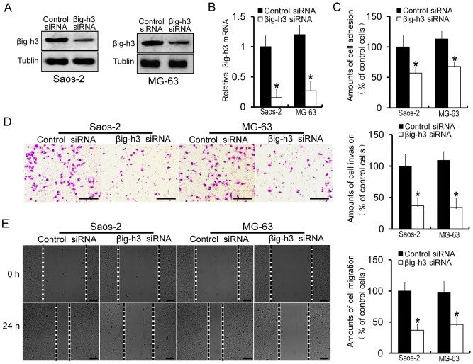
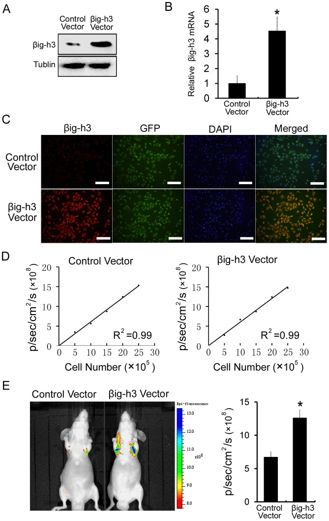
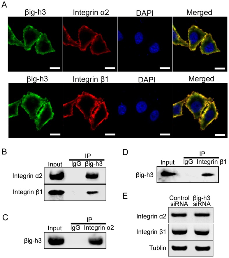
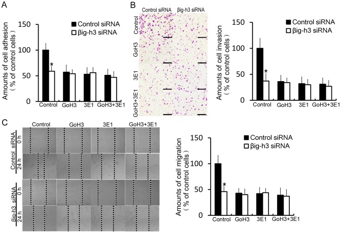
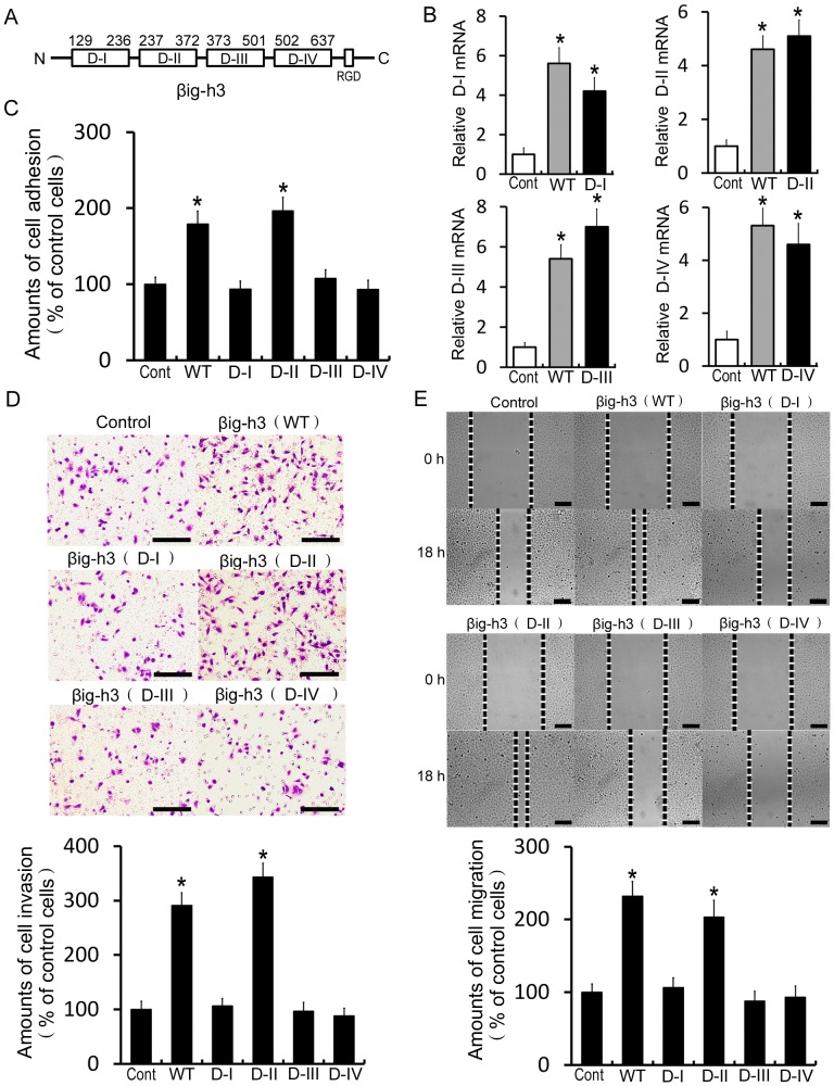
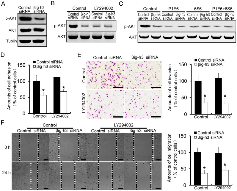
Osteosarcoma, the most common primary bone tumor in children and young adolescents, is characterized by local invasion and distant metastasis. But the detailed mechanisms of osteosarcoma metastasis are not well known. In the present study, we found that βig-h3 promotes metastatic potential of human osteosarcoma cells in vitro and in vivo. Furthermore, βig-h3 co-localized with integrin α2β1 in osteosarcoma cells. But βig-h3 did not change integrin α2β1 expression in Saos-2 cells. Interaction of βig-h3 with integrin α2β1 mediates metastasis of human osteosarcoma cells. The second FAS1 domain of βig-h3 but not the first FAS1 domain, the third FAS1 domain or the fourth FAS1 domain mediates human osteosarcoma cells metastasis, which is the α2β1 integrin-interacting domain. We further demonstrated that PI3K/AKT signaling pathway is involved in βig-h3-induced human osteosarcoma cells metastasis process. Together, these results reveal βig-h3 enhances the metastasis potentials of human osteosarcoma cells via integrin α2β1-mediated PI3K/AKT signal pathways. The discovery of βig-h3-mediated pathway helps us to understand the mechanism of human osteosarcoma metastasis and provides evidence for the possibility that βig-h3 can be a potential therapeutic target for osteosarcoma treatment.


Conflict of interest statement

Competing Interests: The authors have declared that no competing interests exist.

Figures

Figure 1
Figure 1. Downregulation of βig-h3 decreases adhesion, invasion and migration of human osteosarcoma cells in vitro.
(A) Western blot was performed to examine the βig-h3 protein levels in Saos-2 cells and MG63 cells which were transfected with control siRNA or βig-h3 siRNA. (B) Real Time PCR was performed to examine the βig-h3 mRNA levels in Saos-2 cells and MG63 cells which were transfected with control siRNA or βig-h3 siRNA. (C) The amounts of cell adhesion was tested in Saos-2 cells and MG63 cells which were transfected with control siRNA or βig-h3 siRNA. (D) The amounts of cell invasion was tested in Saos-2 cells and MG63 cells which were transfected with control siRNA or βig-h3 siRNA. (E) The amounts of cell migration was tested in Saos-2 cells and MG63 cells which were transfected with control siRNA or βig-h3 siRNA. Control siRNA were used as a negative control. Scale = 100 µm. The adhension assay, invasion assay, and migration assay were adopted as described in Materials and Methods. Values are the means±SE from six independent experiments. *P<0.05 by Student's t test.
Figure 2
Figure 2. βig-h3 promotes metastasis of human osteosarcoma cells in vivo.
(A) Western blot was performed to examine the βig-h3 protein levels in Saos-2 cells which were transfected with control vector or βig-h3 vector. (B) Real Time PCR was performed to examine the βig-h3 mRNA levels in Saos-2 cells which were transfected with control vector or βig-h3 vector. (C) Immunofluorescence was performed to examine the expression levels of GFP and βig-h3 in Saos-2 cells which were transfected with control vector or βig-h3 vector. Scale = 100 µm. (D) A strong correlation exists between the cell number and GFP fluorescence intensity (control vector treated cells, R2 = 0.99; βig-h3 vector treated cells, R2 = 0.99). (E) Representative fluorescence imaging of mice injected with Saos-2 cells stably expressing control vector or βig-h3 vector. Quantification of fluorescence imaging data is shown at the right. Bars represent the mean of triplicate samples; error bars represent standard deviation. Data are representative of three independent experiments. *P<0.05 by Student's t test.
Figure 3
Figure 3. βig-h3 immunoprecipitates with α2β1 integrin in human osteosarcoma cells.
(A) Localization of βig-h3 and integrin α2β1 in Saos-2 cells. Saos-2 cells were double-stained for βig-h3 (green) and integrin α2 and integrin β1 (red). Scale = 2 µm. (B) βig-h3 immunoprecipitation with integrin α2β1. Lysates of Saos-2 cells were subjected to immunoprecipitation with anti- βig-h3 antibody pre-bound coupling gel, integrin α2 and integrin β1 in the immune complexes were detected by western blot analysis. (C) Integrin α2 immunoprecipitation with βig-h3. Lysates of Saos-2 cells were subjected to immunoprecipitation with anti-integrin α2 antibody pre-bound coupling gel, βig-h3 in the immune complexes was detected by Western blot analysis. (D) Integrin β1 immunoprecipitation with βig-h3. Lysates of Saos-2 cells were subjected to immunoprecipitation with anti-integrin β1 antibody pre-bound coupling gel, βig-h3 in the immune complexes was detected by Western blot analysis. Immunoprecipitated with anti-IgG antibody was used as the negative control. (E) Western blot was performed to examine the integrin α2 and integrin β1 protein levels in Saos-2 cells which were transfected with control siRNA or βig-h3 siRNA.
Figure 4
Figure 4. Interaction of βig-h3 with integrin α2β1 mediates metastasis of human osteosarcoma cells.
The amounts of cell adhesion (A), invasion (B) and migration (C) were tested in control siRNA or βig-h3 siRNA transfected Saos-2 cells which were incubated with P1E6 and 6S6, alone or combination. Scale = 100 µm. Bars represent the mean of triplicate samples; error bars represent standard deviation. Data are representative of three independent experiments. *P<0.05 by one-way ANOVA analysis.
Figure 5
Figure 5. The second FAS1 domain of βig-h3 promotes human osteosarcoma cells metastasis.
(A) Schematic representation of the total gene of βig-h3 (WT), the first FAS1domain (D-I),the second FAS1domain (D-II), the third FAS1domain (D-III) and the fourth FAS1domain (D-IV). (B) The total gene of βig-h3 (WT) and its four segments of highly conserved sequence (D-I, D-II, D-III and D-IV) were cloned and transfected into Saos-2 cells. Real Time PCR was used to test the mRNA expression levels of D-I, D-II, D-III and D-IV in Saos-2 cells respectively. βig-h3 (WT) transfected cells were used as positive control. The amounts of cell adhesion (C), invasion (D) and migration (E) were tested in βig-h3 (WT), D-I, D-II, D-III and D-IV transfected Saos-2 cells. Scale = 100 µm. Bars represent the mean of triplicate samples; error bars represent standard deviation. Data are representative of three independent experiments. * p<0.05 by one-way ANOVA analysis.
Figure 6
Figure 6. βig-h3 induces human osteosarcoma cells metastasis by activating PI3K signaling pathway.
(A) Expression levels of phosphorylated AKT (p-AKT) and AKT were analyzed in control siRNA or βig-h3 siRNA transfected Saos-2 cells. (B) Expression levels p-AKT and AKT were analyzed in control siRNA or βig-h3 siRNA transfected Saos-2 cells which were incubated with PI3K inhibitor LY294002. (C) Expression levels of p-AKT and AKT were analyzed in control siRNA or βig-h3 siRNA transfected Saos-2 cells which were incubated with P1E6 and 6S6, alone or combination. The amounts of cell adhesion (D), invasion (E) and migration (F) were tested in control siRNA or βig-h3 siRNA transfected Saos-2 cells which were incubated with LY294002. Scale = 100 µm. Bars represent the mean of triplicate samples; error bars represent standard deviation. Data are representative of three independent experiments. *P<0.05 by Student's t test.

References

    1. Messerschmitt PJ, Garcia RM, Abdul-Karim FW, Greenfield EM, Getty PJ (2009) Osteosarcoma. J Am Acad Orthop Surg 17 (8) 515–527. - PubMed
    1. Kim HJ, Chalmers PN, Morris CD (2010) Pediatric osteogenic sarcoma. Curr Opin Pediatr 22 (1) 61–66. - PubMed
    1. Harris MB, Gieser P, Goorin AM, Ayala A, Shochat SJ, et al. (1998) Treatment of metastatic osteosarcoma at diagnosis: a Pediatric Oncology Group Study. J Clin Oncol 16: 3641–3648. - PubMed
    1. Meyers PS, Heller G, Healey G, Huvos A, Applewhite A, et al. (1993) Osteogenic sarcoma with clinically detectable metastasis at initial presentation. J Clin Oncol 11: 449–453. - PubMed
    1. Woodhouse EC, Chuaqui RF, Liotta LA (1997) General mechanisms of metastasis. Cancer 80 (Suppl. 8) 1529–1537. - PubMed

Publication types

MeSH terms

Substances