. 2014 May;82(5):2037-47.
doi: 10.1128/IAI.01530-14. Epub 2014 Mar 4.

Expression of the effector protein IncD in Chlamydia trachomatis mediates recruitment of the lipid transfer protein CERT and the endoplasmic reticulum-resident protein VAPB to the inclusion membrane

Affiliations

Expression of the effector protein IncD in Chlamydia trachomatis mediates recruitment of the lipid transfer protein CERT and the endoplasmic reticulum-resident protein VAPB to the inclusion membrane

Hervé Agaisse et al. Infect Immun. 2014 May.

Abstract

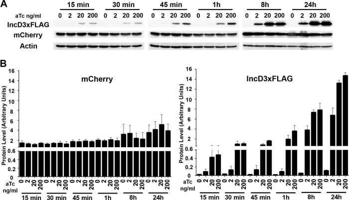
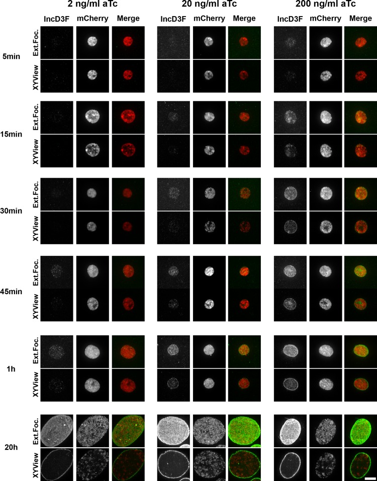
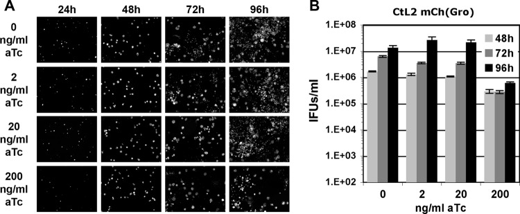
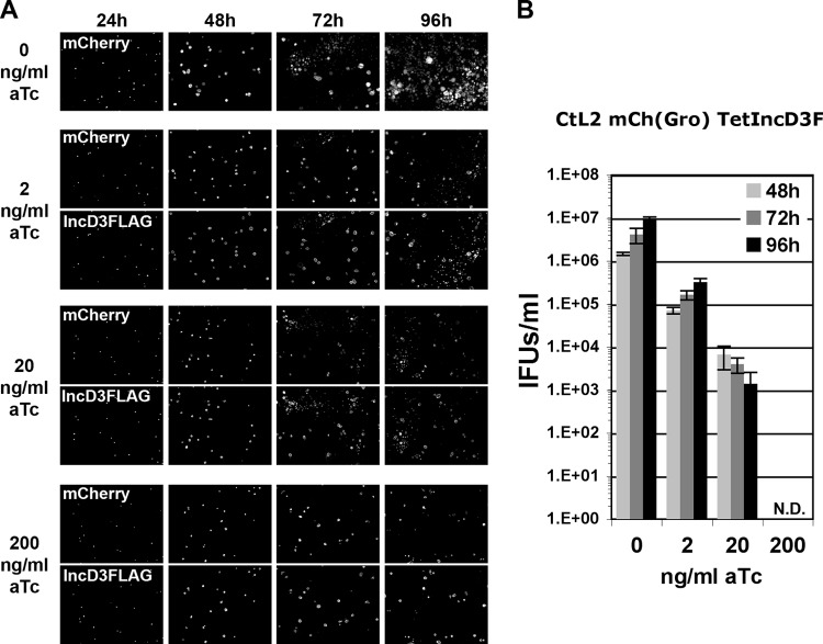
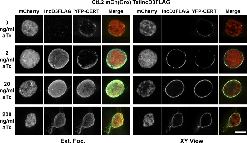
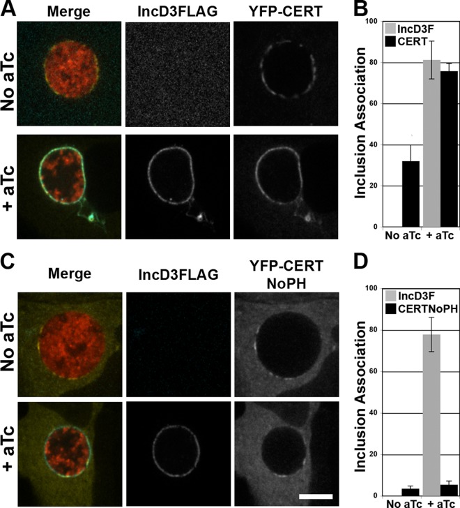
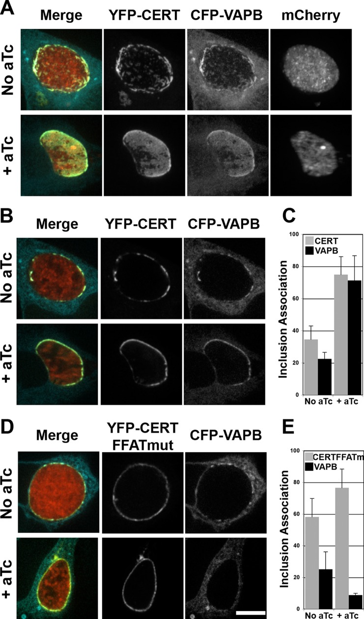
Chlamydia trachomatis is an obligate intracellular human pathogen responsible for ocular and genital infections. To establish its membrane-bound intracellular niche, the inclusion, C. trachomatis relies on a set of effector proteins that are injected into the host cells or inserted into the inclusion membrane. We previously proposed that insertion of the C. trachomatis effector protein IncD into the inclusion membrane contributes to the recruitment of the lipid transfer protein CERT to the inclusion. Due to the genetically intractable status of C. trachomatis at that time, this model of IncD-CERT interaction was inferred from ectopic expression of IncD and CERT in the host cell. In the present study, we investigated the impact of conditionally expressing a FLAG-tagged version of IncD in C. trachomatis. This genetic approach allowed us to establish that IncD-3×FLAG localized to the inclusion membrane and caused a massive recruitment of the lipid transfer protein CERT that relied on the PH domain of CERT. In addition, we showed that the massive IncD-dependent association of CERT with the inclusion led to an increased recruitment of the endoplasmic reticulum (ER)-resident protein VAPB, and we determined that, at the inclusion, CERT-VAPB interaction relied on the FFAT domain of CERT. Altogether, the data presented here show that expression of the C. trachomatis effector protein IncD mediates the recruitment of the lipid transfer protein CERT and the ER-resident protein VAPB to the inclusion.


Figures

FIG 1
FIG 1
Time course of IncD-3×FLAG induction. (A) Immunoblots of cell lysates from cells infected with the CtL2 mCh(Gro) TetIncD3F strain. The cells were infected for 24 h to allow for inclusion formation before addition of 2, 20, or 200 ng/ml aTc. The cells were collected at 15, 30, or 45 min or 1, 8, or 20 h post-aTc induction, and immunoblots of the corresponding lysates were probed using antibodies. (B) Quantification of three independent experiments as presented in panel A. The mCherry (left) and FLAG (right) signal intensities were quantified using Odyssey software and normalized to the actin signal intensity, and protein levels (arbitrary units) were plotted. Please note the split y axis.
FIG 2
FIG 2
IncD-3×FLAG localization during a time course of aTc induction. Confocal micrographs show inclusions of the CtL2 mCh(Gro) TetIncD3F strain. The cells were infected for 24 h to allow for inclusion formation before addition of 2 ng/ml (left panels), 20 ng/ml (middle panels), or 200 ng/ml (right panels) aTc. The cells were fixed at 5, 15, 30, or 45 min or 1 or 20 h post-aTc induction, immunostained with anti-FLAG antibodies, and imaged using a confocal microscope. The same imaging settings were used for all conditions, and the images were not manipulated after acquisition, so the intensities of the signals on the micrographs truly reflect the expression levels of the IncD-3×FLAG construct. For each combination of aTc concentration and time, the left panels correspond to the FLAG signal of the IncD-3×FLAG construct (IncD3F; green) and the middle panels to the bacteria (mCherry; red). Merged images are shown on the right. The top and bottom panels correspond to an extended-focus view combining all the confocal planes (Ext.Foc.) and a single plane crossing the middle of the inclusion (XYView), respectively. Bar, 10 μm.
FIG 3
FIG 3
Growth characteristics of C. trachomatis CtL2 mCh(Gro) strain. (A) Black and white micrographs of confluent HeLa cells infected with a strain of C. trachomatis expressing mCherry under the control of the groESL operon regulatory element [CtL2 mCh(Gro)]. The infected cells were incubated with the indicated concentrations of aTc (0, 2, 20, and 200 ng/ml) at 1 h postinfection and imaged at the indicated times postinfection (24, 48, 72, and 96 h). The channel displaying the mCherry bacteria is shown. (B) Quantification of the numbers of infectious bacteria produced (IFUs/ml) at 48 h, 72 h, and 96 h postinfection (light gray, dark gray, and black bars, respectively) in HeLa cells infected with a strain of C. trachomatis expressing mCherry under the control of the groESL operon regulatory element [CtL2 mCh(Gro)] and incubated in the presence of 0, 2, 20, or 200 ng/ml aTc at 1 h postinfection.
FIG 4
FIG 4
Growth characteristics of the C. trachomatis CtL2 mCh(Gro) TetIncD3F strain. (A) Black and white micrographs of confluent HeLa cells infected with a strain of C. trachomatis expressing mCherry under the control of the groESL operon regulatory element and IncD-3×FLAG under the control of an aTc-inducible promoter [CtL2 mCh(Gro) TetIncD3F]. The infected cells were incubated with the indicated concentrations of aTc (0, 2, 20, and 200 ng/ml) at 1 h postinfection and imaged at the indicated times postinfection (24, 48, 72, and 96 h). The channels displaying the mCherry bacteria and the IncD-3×FLAG construct are shown in the upper (mCherry) and lower (IncD3FLAG) panels, respectively. (B) Quantification of the numbers of infectious bacteria produced (IFUs/ml) at 48 h, 72 h, and 96 h postinfection (light gray, dark gray, and black bars, respectively) in HeLa cells infected with a strain of C. trachomatis expressing mCherry under the control of the groESL operon regulatory element and IncD-3×FLAG under the control of an aTc-inducible promoter [CtL2 mCh(Gro) TetIncD3F] and incubated in the presence of 0, 2, 20, or 200 ng/ml aTc at 1 h postinfection. N.D., none detected.
FIG 5
FIG 5
IncD-3×FLAG expression leads to massive recruitment of CERT to the inclusion membrane. Confocal fluorescence micrographs show inclusions of the CtL2 mCh(Gro) TetIncD3F strain (mCherry; red) in HeLa cells expressing YFP-CERT (YFP-CERT; yellow). At 1 h postinfection, the infected cells were incubated in the absence (0 ng/ml aTc) or presence (2, 20, or 200 ng/ml aTc) of the indicated aTc concentration. The infected cells were fixed at 23 h post-aTc induction, coimmunostained using anti-FLAG (IncD3FLAG; blue), and imaged using a confocal microscope. The same imaging settings were used for all conditions, and the images were not manipulated after acquisition, so the intensities of the signals on the micrographs truly reflect the expression levels of the IncD-3×FLAG construct. The left and right groups of panels correspond to an extended-focus view combining all the confocal planes spanning an entire inclusion (Ext.Foc.) and a single plane crossing the middle of the inclusion (XY View), respectively. Merged images are shown in the right column for each group. Bar, 10 μm.
FIG 6
FIG 6
The massive recruitment of CERT to the inclusion membrane depends on IncD and the PH domain of CERT. (A and C) Confocal fluorescence micrographs of a single plane crossing the middle of inclusions of a C. trachomatis strain, expressing mCherry (red) and IncD3×FLAG (blue) under the control of an aTc-inducible promoter, in HeLa cells expressing wild-type YFP-CERT (YFP-CERT; yellow) (A) or a truncated version lacking the PH domain (YFP-CERT NoPH; yellow) (C). At 1 h postinfection, the infected cells were incubated in the absence (No aTc) or presence (+ aTc) of 20 ng/ml aTc. The cells were fixed at 23 h post-aTc induction, immunostained with anti-FLAG antibodies (IncD3FLAG; blue), and imaged using a confocal microscope. Merged images are shown on the left. Bar, 10 μm. (B and D) Quantifications of the proportions of the inclusion membrane covered by the indicated markers. The quantification method is described in Materials and Methods.
FIG 7
FIG 7
VAPB association with the inclusion membrane is dependent on IncD and the FFAT motif of CERT. (A, B, and D) Confocal fluorescence micrographs of inclusions of a C. trachomatis strain, expressing mCherry (red) and IncD3×FLAG under the control of an aTc-inducible promoter, in HeLa cells coexpressing a CFP-VAPB (blue) construct and either a wild-type YFP-CERT construct (YFP-CERT; yellow) (A and B) or a version displaying a mutation in the FFAT motif (YFP-CERT FFATmut; yellow) (D). At 1 h postinfection, the infected cells were incubated in the absence (No aTc) or presence (+ aTc) of 20 ng/ml aTc. The cells were fixed at 23 h post-aTc induction and imaged using a confocal microscope. An extended-focus view combining all the confocal planes spanning an entire inclusion (A) or views of a single plane crossing the middle of the inclusion (B and D) are shown. Merged images are shown on the left. Bar, 10 μm. (C and E) Quantifications of the proportions of the inclusion membrane covered by the indicated markers. The quantification method is described in Materials and Methods.

References

    1. Friis RR. 1972. Interaction of L cells and Chlamydia psittaci: entry of the parasite and host responses to its development. J. Bacteriol. 110:706–721 - PMC - PubMed
    1. Moulder JW. 1991. Interaction of chlamydiae and host cells in vitro. Microbiol. Rev. 55:143–190 - PMC - PubMed
    1. Bastidas RJ, Elwell CA, Engel JN, Valdivia RH. 2013. Chlamydial intracellular survival strategies. Cold Spring Harb. Perspect. Med. 3:a010256. 10.1101/cshperspect.a010256 - DOI - PMC - PubMed
    1. Bannantine JP, Griffiths RS, Viratyosin W, Brown WJ, Rockey DD. 2000. A secondary structure motif predictive of protein localization to the chlamydial inclusion membrane. Cell. Microbiol. 2:35–47. 10.1046/j.1462-5822.2000.00029.x - DOI - PubMed
    1. Mueller KE, Plano GV, Fields KA. 2014. New frontiers in type III secretion biology: the Chlamydia perspective. Infect. Immun. 82:2–9. 10.1128/IAI.00917-13 - DOI - PMC - PubMed

Publication types

MeSH terms

LinkOut - more resources