. 2014 Mar 3;9(3):e90907.
doi: 10.1371/journal.pone.0090907. eCollection 2014.

Caffeine mediates sustained inactivation of breast cancer-associated myofibroblasts via up-regulation of tumor suppressor genes

Affiliations

Caffeine mediates sustained inactivation of breast cancer-associated myofibroblasts via up-regulation of tumor suppressor genes

Mysoon M Al-Ansari et al. PLoS One. .

Abstract

Background: Active cancer-associated fibroblasts (CAFs) or myofibroblasts play important roles not only in the development and progression of breast carcinomas, but also in their prognosis and treatment. Therefore, targeting these cells through suppressing their supportive procarcinogenic paracrine effects is mandatory for improving the current therapies that are mainly targeting tumor cells. To this end, we investigated the effect of the natural and pharmacologically safe molecule, caffeine, on CAF cells and their various procarcinogenic effects.

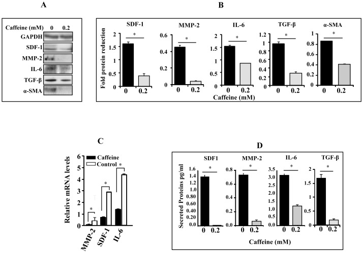
Methodology/principal findings: We have shown here that caffeine up-regulates the tumor suppressor proteins p16, p21, p53 and Cav-1, and reduces the expression/secretion of various cytokines (IL-6, TGF-β, SDF-1 and MMP-2), and down-regulates α-SMA. Furthermore, caffeine suppressed the migratory/invasiveness abilities of CAF cells through PTEN-dependent Akt/Erk1/2 inactivation. Moreover, caffeine reduced the paracrine pro-invasion/-migration effects of CAF cells on breast cancer cells. These results indicate that caffeine can inactivate breast stromal myofibroblasts. This has been confirmed by showing that caffeine also suppresses the paracrine pro-angiogenic effect of CAF cells through down-regulating HIF-1αand its downstream effector VEGF-A. Interestingly, these effects were sustained in absence of caffeine.

Conclusion/significance: The present findings provide a proof of principle that breast cancer myofibroblasts can be inactivated, and thereby caffeine may provide a safe and effective prevention against breast tumor growth/recurrence through inhibition of the procarcinogenic effects of active stromal fibroblasts.


Conflict of interest statement

Competing Interests: The authors have declared that no competing interests exist.

Figures

Figure 1
Figure 1. Caffeine up-regulates the expression of p16, p21, p53 and Cav-1.
Cells were either sham treated or challenged with the indicated caffeine concentrations for 1 µg of proteins were used for immunoblotting analysis using the indicated antibodies. (A) Immunoblots. (B) Histograms show the expression levels of the indicated proteins. The values were determined by densitometry and normalized against GAPDH. Error bars represent means ± S.D.
Figure 2
Figure 2. Caffeine inhibits the expression/secretion of SDF1, IL-6, MMP-2 and TGF-.
CAF-180 cells were either sham-treated or challenged with caffeine for 1 hr. (A) Whole cell lysates were prepared and used for immunoblotting analysis using the indicated antibodies. (B) Histograms show the expression levels of the indicated proteins. The values were determined by densitometry and normalized against GAPDH. (C) Total RNA was prepared and used to assess the level of the indicated genes by qRT-PCR. The obtained values were normalized against β-actin. (D) Secreted levels of proteins were determined in SFCM by ELISA and shown in the histograms. Error bars represent means ± S.D. *: p<0.05.
Figure 3
Figure 3. Caffeine suppresses the invasion/migration of active breast stromal fibroblasts through Akt/Erk1/2 inhibition.
CAF-180 cells were either sham-treated or challenged with caffeine for 1 hr. (A) Cells were cultured on the upper compartments of BioCoat matrigel chambers in the presence of serum-free media. After 18 hr of incubation, cells were stained with Diff-Quick stain, and then were counted. The histograms depict the average numbers of invasive and migrated cells. (B) Whole-cell lysates were prepared and used for immunoblotting analysis using the indicated antibodies. (C) Histograms show the expression levels of the indicated proteins. The values were determined by densitometry and normalized against GAPDH. Error bars represent means ± S.D. *: p<0.05. (D) CAF-180 cells were transfected with PTEN-siRNA or a scrambled sequence, and then cells were either sham-treated of challenged with caffeine for 1 hr. cell lysates were prepared and used for immunoblotting analysis utilizing antibodies against the indicated proteins.
Figure 4
Figure 4. Caffeine inhibits MDA-MB-231 invasion/migration in a paracrine manner.
Serum-free conditioned media were collected from CAF-180 cells either sham-treated or challenged with caffeine (0.2 mM, for 1 hr), SFM as well as complete media (CpM) were also used as control. These media were added separately into the lower compartments of the 24-well BD BioCoat plates. MDA-MB-231 cells (1×10⁴) were seeded onto the upper compartment of the migration (non-coated chambers) and invasion (Matrigel Invasion Chamber) plates and incubated for 18 hr. Average numbers of migrated/invaded cells were depicted in the histograms. Error bars represent means ± S.D. *: p<0.05.
Figure 5
Figure 5. Caffeine effects are persistent.
(A) Cells were either sham-treated (0) or challenged with caffeine (0.2 mM, for 1 hr), and then immediately harvested (1) or reincubated in caffeine-free medium for 24 hr, and then were either harvested (24) or split and reincubated for another 48 hr (split). Subsequently, cell lysates were prepared from all these cells and the levels of various proteins were assessed by immunoblotting. (B) Cells were either sham-treated or challenged with caffeine (0.2 mM) for 1 hr, and then reincubated in caffeine-free medium for 24 hr. Cell lysates were prepared and used for immunoblot analysis. The numbers below the bands represent fold of change as compared to the level at time 0, upon normalization against GAPDH used as internal control. (C) SA-β-gal activity was analyzed. Scale bars represent 50 µm. (D) Histogram shows SA-β-gal labeling index. Error bars represent means ± S.D.
Figure 6
Figure 6. Caffeine inhibits VEGF-A expression/secretion in stromal fibroblasts and their pro-angiogenic effects in vitro.
CAF-180 cells were either sham treated or challenged with caffeine for 1 hr. (A) Whole-cell lysates were prepared and 50 µg of proteins were used for immunoblotting analysis using the indicated antibodies (upper panel). The histograms show the expression levels of the indicated proteins (lower panels). (B) Total RNA was prepared and used to assess the level of VEGF-A by qRT-PCR. The obtained values were normalized against β-actin. (C) CAF-180 cells were treated with caffeine for 1 hr. Non-treated cells were cultured under the same conditions and were used as control. Secreted levels of proteins were determined by ELISA and shown in the histogram. (D) Cells were either sham-treated or challenged with caffeine (0.2 mM) for 1 hr, and then caffeine was removed and cells were washed twice with PBS and were subsequently reincubated in SFM for 24 hr to generate SFCM that have been used to treat HUVEC cells previously plated on matrigel (96-well plate), then incubated at 37°C for 18 hr. (E) Histogram shows the average number of formed cavities. Error bars represent means ± S.D. *: p<0.05.

References

    1. Jemal A, Bray F (2011) Center MM, Ferlay J, Ward E, et al (2011) Global cancer statistics. Ca Cancer J Clin 61: 69–90. - PubMed
    1. Aboussekhra A (2011) Role of cancer-associated fibroblasts in breast cancer development and prognosis. Int J Dev Biol 55: 841–849. - PubMed
    1. Franco OE, Shaw AK, Strand DW, Hayward SW (2009) Cancer associated fibroblasts in cancer pathogenesis. Semin Cell Dev Biol 21: 33–39. - PMC - PubMed
    1. Shimoda M, Mellody KT, Orimo A (2009) Carcinoma-associated fibroblasts are a rate-limiting determinant for tumour progression. Semin Cell Dev Biol 21: 19–25. - PMC - PubMed
    1. Orimo A, Gupta PB, Sgroi DC, Arenzana-Seisdedos F, Delaunay T, et al. (2005) Stromal fibroblasts present in invasive human breast carcinomas promote tumor growth and angiogenesis through elevated SDF-1/CXCL12 secretion. Cell 121: 335–348. - PubMed

Publication types

MeSH terms