

Review
doi: 10.1161/JAHA.113.000582.
Epicardial and perivascular adipose tissues and their influence on cardiovascular disease: basic mechanisms and clinical associations
Affiliations
- PMID: 24595191
- PMCID: PMC4187500
- DOI: 10.1161/JAHA.113.000582
Item in Clipboard
Review
Epicardial and perivascular adipose tissues and their influence on cardiovascular disease: basic mechanisms and clinical associations
J Am Heart Assoc.
.
No abstract available
Keywords: adipose; atherosclerosis; epicardial; obesity; perivascular.
Figures

Anatomy and nomenclature of commonly studied perivascular adipose depots. (1) Paracardial adipose tissue (PAT) is the fat that surrounds the parietal pericardium; it is also referred to as “mediastinal fat” or “thoracic” fat. Recent human data suggest that this fat may be “beige” in morphology―that is, having features of both white and brown adipose tissue.(2013) Measurement of paracardial fat volume is often used in imaging studies and may or may not be specifically differentiated from the underlying epicardial fat. (2) Epicardial adipose tissue (EAT) is encased by the visceral pericardium, lying directly adjacent to the myocardium and surrounding the coronary arteries. Here, it is shown surrounding the right coronary artery (RCA) and left anterior descending coronary artery (LAD). Human EAT is morphologically similar to white adipose tissue.(2013)
Pericardial fat has been defined as the combination of paracardial and epicardial fat.(2005) (3) Thoracic periaortic fat surrounds the thoracic aorta in humans and rodents. In rodents, it is morphologically and functionally identical to brown adipose tissue; whether this is the case in humans remains unknown.(2011)–(2012) (4) Abdominal periaortic fat surrounds the abdominal aorta and has features of white adipose tissue in rodents and humans.(2009),(2005) (5) Small artery fat, or that surrounding the mesenteric arteries and smaller arteries, usually has features of white adipose. Fat surrounding small arteries (ie, resistance vessels) may play a role in regulating vascular tone and flow of metabolites/nutrients, as originally proposed by Yudkin.(2009)–(2005)
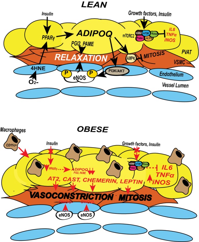
Signaling pathways that mediate the paracrine effects of PVAT. Adiponectin (ADIPOQ) has been shown to mediate many of the beneficial effects of PVAT. ADIPOQ is more highly expressed in lean conditions (top) than in obese conditions (bottom). ADIPOQ causes arterial vasodilation by promoting local eNOS activity in 2 ways: stimulation of AKT‐dependent phosphorylation and subsequent increased eNOS coupling, and by increasing BH4 cofactor availability.(2013) ROS byproducts generated by dysfunctional endothelium (4HNE) feedback to activate PPARγ in adjacent PVAT, thereby stimulating transcription of ADIPOQ and other downstream adipogenic genes. ADIPOQ also promotes endothelium‐independent vasodilation by activation of VSMC potassium channels (not shown). ADIPOQ released by healthy PVAT prevents neointimal hyperplasia by suppressing mitosis of VSMCs in an AMPK‐dependent mechanism(2009) (top). Lipids such as palmitic acid methyl ester (PAME) and prostacyclin (PGI2) have also been shown to stimulate endothelium‐independent vasodilation via direct activation of VSMC potassium channels and PGI2 receptor activation (top).(2012),(2011) Finally, in PVAT from lean mice, unknown growth factor signals converge on mTORC2, resulting in Akt activation and inhibition of inflammatory cytokine production by adipocytes; loss of mTORC2 signaling in Rictor‐deficient mice results in increased inflammation and increased vasoconstriction of underlying VSMC (top).(2013) In obesity, these pathways may be compromised, or new pathological mechanisms may emerge (bottom). Obesity stimulates inflammation and macrophage infiltration, which increases local cytokine and ROS production (bottom). The resultant impairment of insulin signaling contributes to a reduction in beneficial adipokine expression (ADIPOQ, PAME, PGI2), uncoupling of eNOS, and increased expression of pathological vasoconstricting factors (angiotensin 2 [AT2], chemerin, and calpastatin).(2013) PVAT from obese patients may also release factors that stimulate mitosis (eg, leptin, PDGF) of VSMCs by overwhelming local beneficial adipokine effects.(2009),(2011) AMPK indicates adenosine monophosphate kinase; BH4, tetrahydrobiopterin; eNOS, endothelial nitric oxide synthetase; 4HNE, 4‐hydroxynonenal; IL‐6, interleukin 6; iNOS, inducible nitric oxide synthetase; mLST8, MTOR associated protein, LST8; mTORC2, mammalian target of rapamycin complex 2; ONOO−, peroxynitrite; PDGF, platelet‐derived growth factor; PI3K/AKT, Phosphoinositide 3‐kinase; PPARγ, peroxisome proliferator–activated receptor gamma; PVAT, perivascular adipose tissue; ROS, reactive oxygen species; TNFα, tumor necrosis factor alpha; VSMC, vascular smooth muscle cell.
References
-
- Fox CS, Massaro JM, Hoffmann U, Pou KM, Maurovich‐Horvat P, Liu CY, Vasan RS, Murabito JM, Meigs JB, Cupples LA, D'Agostino RB, Sr, O'Donnell CJ. Abdominal visceral and subcutaneous adipose tissue compartments: association with metabolic risk factors in the Framingham Heart Study. Circulation. 2007; 116:39-48 - PubMed
-
- Van Gaal LF, Mertens IL, De Block CE. Mechanisms linking obesity with cardiovascular disease. Nature. 2006; 444:875-880 - PubMed
-
- Litwin SE. Normal weight obesity: is bigger really badder? Circ Cardiovasc Imaging. 2012; 5:286-288 - PubMed
-
- Iacobellis G, Assael F, Ribaudo MC, Zappaterreno A, Alessi G, Di Mario U, Leonetti F. Epicardial fat from echocardiography: a new method for visceral adipose tissue prediction. Obes Res. 2003; 11:304-310 - PubMed
Publication types
MeSH terms
Substances
Grants and funding
LinkOut - more resources
Full Text Sources
Other Literature Sources
Medical
