Review
. 2014 Mar 3;15(3):3768-83.
doi: 10.3390/ijms15033768.

Three-dimensional structural aspects of protein-polysaccharide interactions

Affiliations
Review

Three-dimensional structural aspects of protein-polysaccharide interactions

Masamichi Nagae et al. Int J Mol Sci. .

Abstract

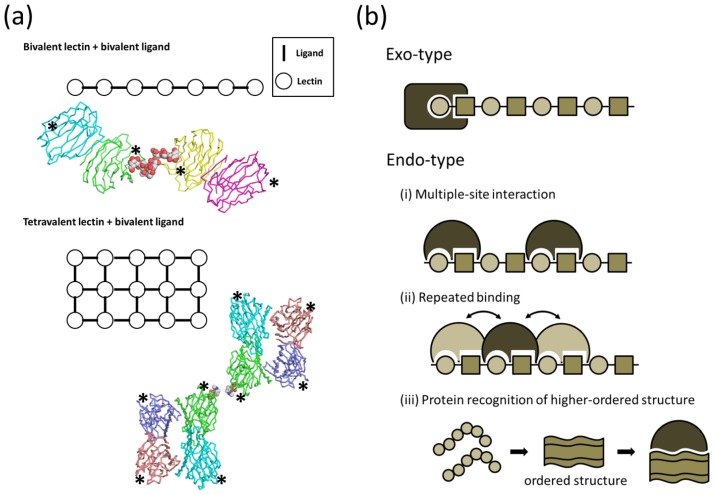
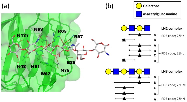
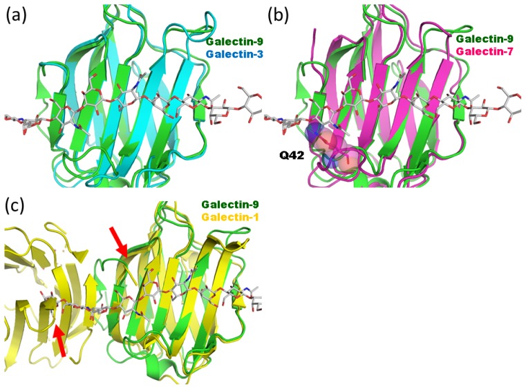
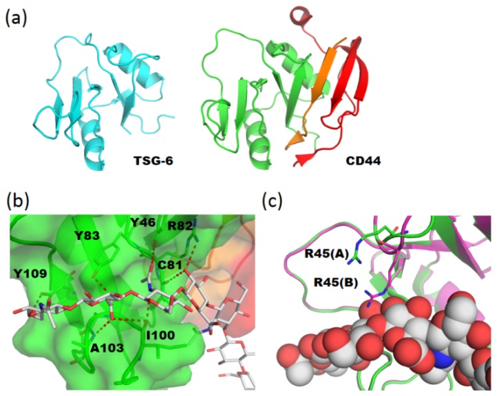
Linear polysaccharides are typically composed of repeating mono- or disaccharide units and are ubiquitous among living organisms. Polysaccharide diversity arises from chain-length variation, branching, and additional modifications. Structural diversity is associated with various physiological functions, which are often regulated by cognate polysaccharide-binding proteins. Proteins that interact with linear polysaccharides have been identified or developed, such as galectins and polysaccharide-specific antibodies, respectively. Currently, data is accumulating on the three-dimensional structure of polysaccharide-binding proteins. These proteins are classified into two types: exo-type and endo-type. The former group specifically interacts with the terminal units of polysaccharides, whereas the latter with internal units. In this review, we describe the structural aspects of exo-type and endo-type protein-polysaccharide interactions. Further, we discuss the structural basis for affinity and specificity enhancement in the face of inherently weak binding interactions.


Figures

Figure 1.
Figure 1.
(a) Schematic representations of bivalent lectin-bivalent ligand (type I) and tetravalent lectin-bivalent ligand (type II) complexes. Crystal packings are shown for bivalent galectin-1 dimer in complex with biantennary complex-type glycan (type I, upper panel, PDB code 1SLB), and tetravalent soybean agglutinin tetramer in complex with binantennary complex-type glycan (type II, lower panel, PDB code 1SBE). Protein and carbohydrate molecules are shown in wire and sphere models, respectively, and arbohydrate binding sites are shown with asterisks; (b) Schematic representations of exo-type (upper) and endo-type (lower) interactions. The affinity enhancement strategies of endo-type lectins: (i) multiple-site interaction; (ii) repeated binding; and (iii) protein recognition of ordered/higher-ordered polysaccharide structures.
Figure 2.
Figure 2.
Polylactosamine recognition by galectins. (a) Structure of human galectin-9 N-terminal carbohydrate recognition domain (NCRD)–LacNAc trimers (LN3) complex (PDB code 2ZHN). The protein molecule is shown in semi-transparent surface and ribbon models. The amino-acid residues that interact with carbohydrate residues are shown in rod models. Hydrogen bonds are indicated by red dotted lines; (b) Schematic representation of the interaction modes of LN2 and LN3 complexes. Galactose residues that interact with conserved key residues in galectin-9 NCRD are shown as black triangles. Carbohydrate residues that interact with galectin-9 NCRD are indicated by double head arrows.
Figure 3.
Figure 3.
Structural comparison of galectin CRDs. (a) Structural superpositions between hypothetical galectin-9 NCRD-LN5 complex (green) and human galectin-3 CRD (cyan; PDB code 1A3K) [17]; (b) Structural comparison with human galectin-7 CRD (magenta; PDB code 1BKZ) [18]. The Gln42 side chain, which collides with LN5, is shown in rod model; (c) Structural comparison with human dimeric galectin-1 CRD (yellow; PDB code 3OYW) [23]. Possible steric clashes are indicated with red arrows.
Figure 4.
Figure 4.
(a) Overall structures of tumor necrosis factor stimulating gene-6 (TSG-6) (PDB code 1O7B, [35], left panel) and CD44 (PDB code; 2JCQ, [31], right panel) Link modules. The extended N- and C-terminal regions of CD44 are colored in orange and red, respectively; (b) Close up view of CD44–HA8 complex (PDB code 2JCQ). The protein molecule is shown in semi-transparent surface and ribbon models. The amino-acid residues that interact with carbohydrate residues are shown in rod models. Hydrogen bonds are indicated by red dotted lines; (c) Structural superposition of two crystal forms of CD44-HA8 complex (crystal form A; green, 2JCQ, crystal form B; magenta, 2JCR). HA8 molecule is shown in sphere model.
Figure 5.
Figure 5.
Interaction between cellulose-binding module (CBM) and β1–3 glucan. (a) Close-up view of the ligand-binding site of TmCBM4-2 in complex with laminariheptaose (PDB code 1GUI) [39]. Protein molecule is shown in semi-transparent surface and ribbon models. Carbohydrate is shown in rod model; (b) Close-up view of the ligand-binding site of BhCBM6 in complex with laminarihexaose (PDB code 1W9W) [40]; (c) Close-up view of the ligand-binding site of βGRP-N in complex with laminarihexaose (PDB code 3AQX) [43]; Hydrogen bonds are indicated by red dotted lines; (d) Structural comparison of three CBMs, TmCBM4-2 (left), BhCBM6 (middle) and βGRP-N (right). Protein and carbohydrate are shown in ribbon and sphere models, respectively.
Figure 6.
Figure 6.
Structural analysis of anti-polysialic acid antibody and oligosialic acid. (a) Schematic representation of a single-chain variable fragment of mAb735 (scFv735) in complex with octasialic acid is shown in the upper panel (PDB code 3WBD) [52]. Two proteins and one carbohydrate in the asymmetric unit are shown in the surface and sphere models, respectively (lower panel); (b) Close-up view of the carbohydrate-recognition site. Direct and water-mediated interactions between scFv735 and trisialic acid are shown in the left and right panels, respectively. Hydrogen bonds are shown as red dotted lines. Water molecules that bridge scFv735 and trisialic acid are shown as red spheres.

Similar articles

Cited by

References

    1. Lee R.T., Lee Y.C. Affinity enhancement by multivalent lectin-carbohydrate interaction. Glycoconj. J. 2000;17:543–551. - PubMed
    1. Fred Brewer C. Binding and cross-linking properties of galectins. Biochim. Biophys. Acta. 2002;1572:255–262. - PubMed
    1. Bourne Y., Bolgiano B., Liao D.I., Strecker G., Cantau P., Herzberg O., Feizi T., Cambillau C. Crosslinking of mammalian lectin (galectin-1) by complex biantennary saccharides. Nat. Struct. Biol. 1994;1:863–870. - PubMed
    1. Gupta D., Bhattacharyya L., Fant J., Macaluso F., Sabesan S., Brewer C.F. Observation of unique cross-linked lattices between multiantennary carbohydrates and soybean lectin Presence of pseudo-2-fold axes of symmetry in complex type carbohydrates. Biochemistry. 1994;33:7495– 7504. - PubMed
    1. Olsen L.R., Dessen A., Gupta D., Sabesan S., Sacchettini J.C., Brewer C.F. X-ray crystallographic studies of unique cross-linked lattices between four isomeric biantennary oligosaccharides and soybean agglutinin. Biochemistry. 1997;36:15073–15080. - PubMed

LinkOut - more resources