. 2014 Mar 3;15(3):3816-33.
doi: 10.3390/ijms15033816.

Role of A20 in cIAP-2 protection against tumor necrosis factor α (TNF-α)-mediated apoptosis in endothelial cells

Affiliations

Role of A20 in cIAP-2 protection against tumor necrosis factor α (TNF-α)-mediated apoptosis in endothelial cells

Shuzhen Guo et al. Int J Mol Sci. .

Abstract

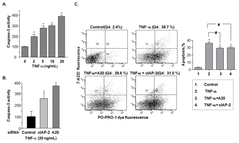
Tumor necrosis factor α (TNF-α) influences endothelial cell viability by altering the regulatory molecules involved in induction or suppression of apoptosis. However, the underlying mechanisms are still not completely understood. In this study, we demonstrated that A20 (also known as TNFAIP3, tumor necrosis factor α-induced protein 3, and an anti-apoptotic protein) regulates the inhibitor of apoptosis protein-2 (cIAP-2) expression upon TNF-α induction in endothelial cells. Inhibition of A20 expression by its siRNA resulted in attenuating expression of TNF-α-induced cIAP-2, yet not cIAP-1 or XIAP. A20-induced cIAP-2 expression can be blocked by the inhibition of phosphatidyl inositol-3 kinase (PI3-K), but not nuclear factor (NF)-κB, while concomitantly increasing the number of endothelial apoptotic cells and caspase 3 activation. Moreover, TNF-α-mediated induction of apoptosis was enhanced by A20 inhibition, which could be rescued by cIAP-2. Taken together, these results identify A20 as a cytoprotective factor involved in cIAP-2 inhibitory pathway of TNF-α-induced apoptosis. This is consistent with the idea that endothelial cell viability is dependent on interactions between inducers and suppressors of apoptosis, susceptible to modulation by TNF-α.


Figures

Figure 1.
Figure 1.
A20 expression is upregulated by tumor necrosis factor α (TNF-α) in endothelial cells. (A) Quantitative PCR analysis of A20 mRNA expression in both human aortic endothelial cells (HAECs) and bovine aortic endothelial cells (BAECs) stimulated with TNF-α (20 ng/mL) for 4 h. The data is presented from triplet tests as means ± SD. * p < 0.05; (B) Expression of A20 was evaluated by immunoblot analysis in BAEC cells before and after 20 ng/mL TNF-α treatment for 4 h. Data shown is a representative blot of three experiments performed; and (C) The effect of TNF-α stimulation on A20 promoter activity was examined by luciferase activity assay in HEK 293 cells. Analysis was performed after 4 h treatment of different doses of TNF-α. Results are expressed as luciferase activity from triplicate tests, and are presented as means ± SD. Statistical significance was determined as * p < 0.05 compared with control.
Figure 2.
Figure 2.
A20 induces the expression of cellular inhibitor of apoptosis protein (cIAP)-2 but not cIAP-1. (A) Quantitative PCR analysis for cIAP-2 mRNA expression was performed in BAEC cells. The data is presented from triplicate tests as means ± SD. * p < 0.05; (B) Immunoblot analysis comparing cIAP-2, cIAP-1 and XIAP in control and A20 overexpressing BAEC and HAEC cells. Data shown is a representative blot of three experiments performed (left) and the related statistical graph (right). * p < 0.05; (C) Quantitative PCR analysis for cIAP-2 mRNA levels (left) and immunoblot analysis for cIAP-2 protein expression (right) was performed in BAEC cells, after knocking down A20 expression by two different siRNA sequences targeted to A20. The data is presented from triplicate tests as means ± SD. * p < 0.05; and (D) Effect of A20 on cIAP-2 promoter activity was measured by luciferase assay in HEK293 cells transfected with A20 or control plasmids. The data is presented from triplicate tests as means ± SD. Statistical significance was determined as * p < 0.05 compared with control.
Figure 3.
Figure 3.
cIAP-2 is upregulated by TNF-α-induced A20. (A) Northern blot analysis of cIAP-2 mRNA levels after stimulation with TNF-α (20 ng/mL), FGF2 (25 ng/mL), VEGF (25 ng/mL), IGF-1 (50 ng/mL), PDGF-AA (20 ng/mL), PDGF-BB (20 ng/mL), PDGF-AB (20 ng/mL), HGF (20 ng/mL) and IL1-β (5 ng/mL) in BAECs; (B) Northern blot showed TNF-α-induced cIAP-2 mRNA expression demonstrated as a dose-(upper) and time-(lower) dependent manner; (C) Quantitative-PCR analysis of cIAP-2 mRNA after stimulation with TNF-α (20 ng/mL), VEGF (25 ng/mL), IGF-1 (50 ng/mL) and IL1-β (5 ng/mL) for 4 h in HAECs. Results are expressed as fold change of control from triplicate tests, and are presented as means ± SD. * p < 0.05; (D) Quantitative-PCR analysis of cIAP-1, cIAP-2 and XIAP mRNA levels after stimulation with TNF-α (20 ng/mL) for 4 h in HAECs. Results are expressed as fold changes of control from triplicate tests, and are presented as means ± SD. * p < 0.05; (E) Effect of TNF-α stimulation on cIAP-2 and cIAP-1 promoter activity by luciferase activity assay. Results are expressed as fold change to control from triplicate tests, and are presented as means ± SD. * p < 0.05; and (F) Targeted knockdown of A20 expression by A20 siRNAs after TNF-α (20 ng/mL) stimulation of cIAP-2 protein expression analyzed by immunoblot (left) and mRNA levels by quantitative PCR (right). Results are expressed as fold change to control from triplicate tests, and are presented as means ± SD. * p < 0.05.
Figure 3.
Figure 3.
cIAP-2 is upregulated by TNF-α-induced A20. (A) Northern blot analysis of cIAP-2 mRNA levels after stimulation with TNF-α (20 ng/mL), FGF2 (25 ng/mL), VEGF (25 ng/mL), IGF-1 (50 ng/mL), PDGF-AA (20 ng/mL), PDGF-BB (20 ng/mL), PDGF-AB (20 ng/mL), HGF (20 ng/mL) and IL1-β (5 ng/mL) in BAECs; (B) Northern blot showed TNF-α-induced cIAP-2 mRNA expression demonstrated as a dose-(upper) and time-(lower) dependent manner; (C) Quantitative-PCR analysis of cIAP-2 mRNA after stimulation with TNF-α (20 ng/mL), VEGF (25 ng/mL), IGF-1 (50 ng/mL) and IL1-β (5 ng/mL) for 4 h in HAECs. Results are expressed as fold change of control from triplicate tests, and are presented as means ± SD. * p < 0.05; (D) Quantitative-PCR analysis of cIAP-1, cIAP-2 and XIAP mRNA levels after stimulation with TNF-α (20 ng/mL) for 4 h in HAECs. Results are expressed as fold changes of control from triplicate tests, and are presented as means ± SD. * p < 0.05; (E) Effect of TNF-α stimulation on cIAP-2 and cIAP-1 promoter activity by luciferase activity assay. Results are expressed as fold change to control from triplicate tests, and are presented as means ± SD. * p < 0.05; and (F) Targeted knockdown of A20 expression by A20 siRNAs after TNF-α (20 ng/mL) stimulation of cIAP-2 protein expression analyzed by immunoblot (left) and mRNA levels by quantitative PCR (right). Results are expressed as fold change to control from triplicate tests, and are presented as means ± SD. * p < 0.05.
Figure 4.
Figure 4.
The PI3-K signaling pathway is involved in A20-mediated TNF-α-induced cIAP-2 expression. (A) Quantitative-PCR analysis of cIAP-2 mRNA levels in HAECs pre-treated with LY294002 (25 μM, PI3-K inhibitor), U0126 (10 μM, ERK1/2 inhibitor), SB203580 (20 μM, p38 inhibitor), and SP600125 (20 μM, JNK inhibitor), respectively for 1 h. Results are expressed as fold change to control from triplicate tests, and are presented as means ± SD. * p < 0.05; (B) Immunoblot analysis in A20 o/e HAECs pre-treated with either wedelolactone (20 μM, IKKα/β inhibitor) or LY294002 (25 μM, PI3-K inhibitor) for 1 h and then immunoblotted for A20, cIAP-2 and vinculin. Representative blot is shown; and (C) Immunoblot analysis in HAECs stimulated with TNF-α (20 ng/mL) and together with either (20 μM, IKKα/β inhibitor) or LY294002 (25 μM, PI3-K inhibitor). A20, cIAP-1 and β-actin were blotted. Representative blot is shown.
Figure 5.
Figure 5.
A20-mediated cIAP-2 is important in endothelial cell resistance to TNF-α-induced apoptosis (caspase3 activity). (A) Caspase-3 activity was analyzed in the BAEC cell lysates treated with various concentrations of TNF-α by means of a colorimetric assay calculated as pmol p-nitroaniline released per min per μg cellular protein. Colorimetric readings were performed in a plate reader at an optical density of 405 nm. The data is presented as percentage of control from triplicate tests as means ± SD. * p < 0.05; (B) Caspase-3 activity was analyzed in TNF-α-stimulated BAEC cells transfected with siRNAs targeted to cIAP-2 and A20 siRNA to knockdown expression. The data is presented as percentage of control from triplicate tests as means ± SD. * p < 0.05; (C) Flow cytometry analysis of endothelial cells stained with PO-PRO-1 dye and 7-AAD to examine the effect of A20 and cIAP-2 overexpression (induced by transfection) on TNF-α-induced apoptotic cells. Representative flow cytometry plots (left) and statistic graph (means ± SD) (right) were shown. * p < 0.05, vs. control cells; # p < 0.05, vs. TNF-α-stimulated control cells; and (D) Flow cytometry analysis of endothelial cells stained with PO-PRO-1 dye and 7-AAD to examine the effect of knockdown of either A20 or cIAP-2 by siRNAs on TNF-α-induced apoptotic cells, or transfection with siRNAs targeted to A20 or cIAP-2 and/or plasmids containing full length cDNA of cIAP-2 or A20 respectively to determine if TNF-α-induced apoptotis could be rescued. Representative flow cytometry plots (left) and statistic graph (means ± SD) (right) were shown. * p < 0.05, vs. control siRNA group; ‡ p < 0.05, vs. control siRNA+ TNF-α group; # p < 0.05, vs. cIAP-2 siRNA+ TNF-α group; † p < 0.05, vs. A20 siRNA+ TNF-α group.
Figure 5.
Figure 5.
A20-mediated cIAP-2 is important in endothelial cell resistance to TNF-α-induced apoptosis (caspase3 activity). (A) Caspase-3 activity was analyzed in the BAEC cell lysates treated with various concentrations of TNF-α by means of a colorimetric assay calculated as pmol p-nitroaniline released per min per μg cellular protein. Colorimetric readings were performed in a plate reader at an optical density of 405 nm. The data is presented as percentage of control from triplicate tests as means ± SD. * p < 0.05; (B) Caspase-3 activity was analyzed in TNF-α-stimulated BAEC cells transfected with siRNAs targeted to cIAP-2 and A20 siRNA to knockdown expression. The data is presented as percentage of control from triplicate tests as means ± SD. * p < 0.05; (C) Flow cytometry analysis of endothelial cells stained with PO-PRO-1 dye and 7-AAD to examine the effect of A20 and cIAP-2 overexpression (induced by transfection) on TNF-α-induced apoptotic cells. Representative flow cytometry plots (left) and statistic graph (means ± SD) (right) were shown. * p < 0.05, vs. control cells; # p < 0.05, vs. TNF-α-stimulated control cells; and (D) Flow cytometry analysis of endothelial cells stained with PO-PRO-1 dye and 7-AAD to examine the effect of knockdown of either A20 or cIAP-2 by siRNAs on TNF-α-induced apoptotic cells, or transfection with siRNAs targeted to A20 or cIAP-2 and/or plasmids containing full length cDNA of cIAP-2 or A20 respectively to determine if TNF-α-induced apoptotis could be rescued. Representative flow cytometry plots (left) and statistic graph (means ± SD) (right) were shown. * p < 0.05, vs. control siRNA group; ‡ p < 0.05, vs. control siRNA+ TNF-α group; # p < 0.05, vs. cIAP-2 siRNA+ TNF-α group; † p < 0.05, vs. A20 siRNA+ TNF-α group.

Similar articles

Cited by

References

    1. Winn R.K., Harlan J.M. The role of endothelial cell apoptosis in inflammatory and immune diseases. J. Thromb. Haemost. 2005;3:1815–1824. - PubMed
    1. Badrichani A.Z., Stroka D.M., Bilbao G., Curiel D.T., Bach F.H., Ferran C. Bcl-2 and bcl-xl serve an anti-inflammatory function in endothelial cells through inhibition of NF-κB. J. Clin. Investig. 1999;103:543–553. - PMC - PubMed
    1. He K.L., Ting A.T. A20 inhibits tumor necrosis factor (TNF) α-induced apoptosis by disrupting recruitment of tradd and rip to the tnf receptor 1 complex in jurkat T cells. Mol. Cell. Biol. 2002;22:6034–6045. - PMC - PubMed
    1. Karsan A., Yee E., Harlan J.M. Endothelial cell death induced by tumor necrosis factor-α is inhibited by the bcl-2 family member a1. J. Biol. Chem. 1996;271:27201–27204. - PubMed
    1. Stehlik C., de Martin R., Kumabashiri I., Schmid J.A., Binder B.R., Lipp J. Nuclear factor (NF)-κB-regulated X-chromosome-linked iap gene expression protects endothelial cells from tumor necrosis factor α-induced apoptosis. J. Exp. Med. 1998;188:211–216. - PMC - PubMed

Publication types

MeSH terms

Substances

LinkOut - more resources