Review
. 2014 Mar;141(6):1184-96.
doi: 10.1242/dev.098285.

Shared signaling systems in myeloid cell-mediated muscle regeneration

Affiliations
Review

Shared signaling systems in myeloid cell-mediated muscle regeneration

James G Tidball et al. Development. 2014 Mar.

Abstract

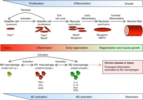
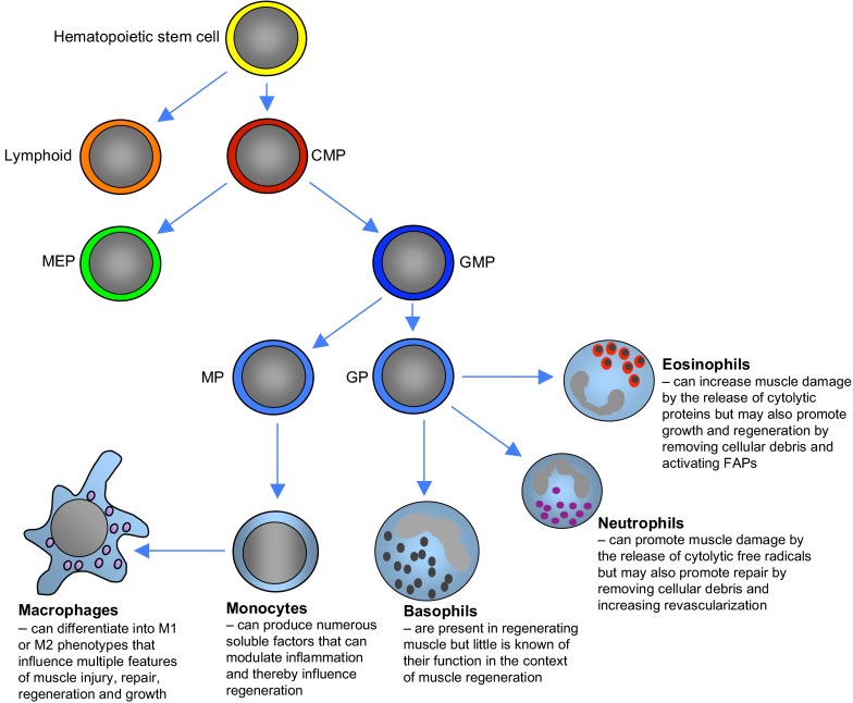
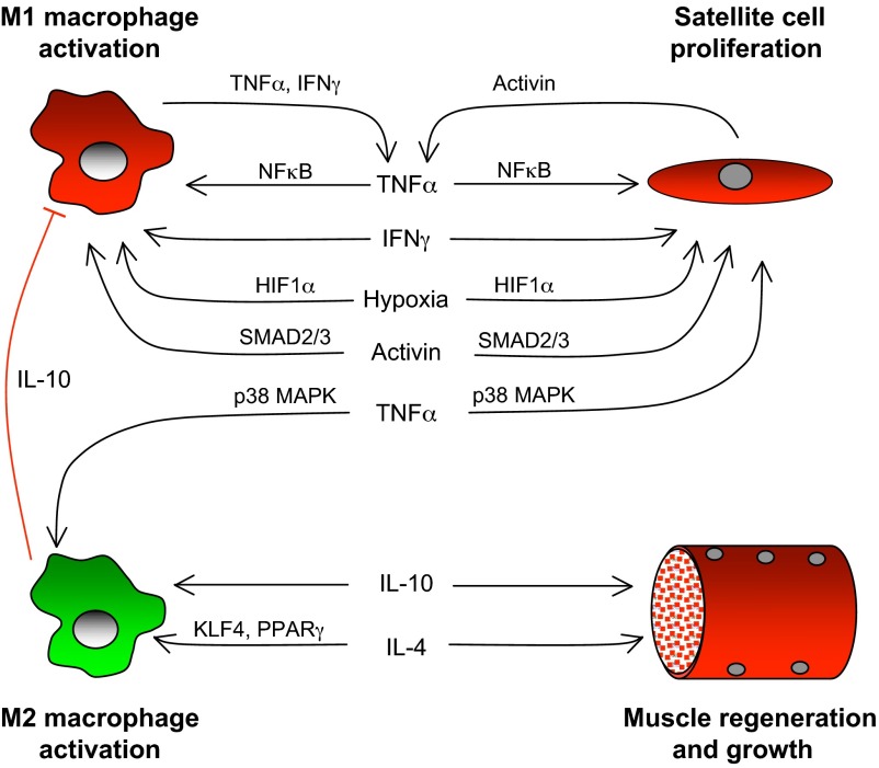
Much of the focus in muscle regeneration has been placed on the identification and delivery of stem cells to promote regenerative capacity. As those efforts have advanced, we have learned that complex features of the microenvironment in which regeneration occurs can determine success or failure. The immune system is an important contributor to that complexity and can determine the extent to which muscle regeneration succeeds. Immune cells of the myeloid lineage play major regulatory roles in tissue regeneration through two general, inductive mechanisms: instructive mechanisms that act directly on muscle cells; and permissive mechanisms that act indirectly to influence regeneration by modulating angiogenesis and fibrosis. In this article, recent discoveries that identify inductive actions of specific populations of myeloid cells on muscle regeneration are presented, with an emphasis on how processes in muscle and myeloid cells are co-regulated.

Keywords: Macrophage phenotype; Muscle regeneration; Signaling systems.


Figures

Fig. 1.
Fig. 1.
Changes in macrophage phenotype and stages of myogenesis in regenerative muscle following injury. During the initial stages of inflammation, Pax7+ satellite cells are activated, proliferate and begin expression of MyoD, initiating transcription of muscle-specific genes necessary for early differentiation. These events coincide with the activation of macrophages from the M0 phenotype to the CD68high/CD206- M1 phenotype, when they express elevated levels of TNFα, IFNγ and iNOS. As myogenesis proceeds, some activated satellite cells return to quiescence and renew the satellite cell reserve population, while others exit the cell cycle to undergo further differentiation. Those post-mitotic myocytes display a shift in gene expression that enables their fusion to form multinucleated myotubes that are able to undergo terminal differentiation. The progress of muscle cells into early regenerative stages coincides with a shift of macrophages to an M2 phenotype that expresses IL-10, IL-4, arginase 1 (Arg1) and TGFβ. As muscle regeneration and growth proceed to restore normal homeostasis, the inflammatory response is slowly resolved. However, in the case of chronic injury or repeated acute injuries, prolonged activity of M2 macrophages can exacerbate muscle dysfunction by driving the excessive accumulation of connective tissue, which is attributable to prolonged production of the pro-fibrotic cytokine TGFβ and to increased metabolism of arginine by Arg1.
Fig. 2.
Fig. 2.
Lineage of myeloid cells that can influence muscle regeneration. Common myeloid progenitors (CMPs) differentiate from multipotent hematopoietic stem cells to give rise to the numerous lines of myeloid cells. CMPs first differentiate into megakaryocyte and erythroid progenitor cells (MEPs) or granulocyte and macrophage progenitor cells (GMPs), with the latter lineage giving rise to the myeloid cell populations that have been most clearly associated with muscle regeneration. GMPs in circulating populations differentiate into either monocyte precursors (MPs) or granulocyte precursors (GPs), which can then undergo terminal differentiation into committed populations that can influence muscle injury, growth or regeneration. MPs give rise to monocytes that release a vast number of soluble factors that modulate the inflammatory response. Monocytes undergo further differentiation to become macrophages, the best characterized population of myeloid cells that influence muscle regeneration. GPs give rise to other populations of myeloid cells that can also influence regeneration. Eosinophils that derive from GPs can either promote muscle damage or muscle regeneration, which varies according to the injury or disease that initiates the inflammatory response. Neutrophils also play a dual role and have the capacity to either amplify muscle damage or promote repair by contributing to the removal of damaged tissue and increasing revascularization. Basophils, which are also derived from GPs, are present in regenerating muscle, although it is not clear whether they have a specific role in muscle regeneration. FAPs, fibro/adipogenic progenitors.
Fig. 3.
Fig. 3.
Coordination of macrophage-mediated signaling and muscle regeneration. Expression of TNFα and IFNγ is elevated during the proliferative stage of myogenesis during muscle regeneration and can contribute to coordinating the inflammatory and myogenic processes by activating macrophages to the M1 phenotype while increasing satellite cell proliferation and inhibiting differentiation. TNFα activation of NFκB in M1 macrophages can promote these effects by further activating TNFα and IFNγ expression in M1 macrophages. Additional positive feedback for M1 activation and satellite cell proliferation can occur through TNFα activation of activin A expression by muscle cells. Increased expression and release of activin A by muscle cells, which has an autocrine effect on muscle cells, inhibits differentiation and maintains muscle cells in the proliferative stage of myogenesis. The released activin A also promotes TNFα expression in macrophages through a SMAD2/3 signaling pathway, maintaining the M1 phenotype. Finally, hypoxia, which is secondary to vascular damage in the injured tissue, activates the expression of genes under the transcriptional control of HIF1α, which promotes the M1 phenotype in macrophages and drives the proliferation of satellite cells. However, TNFα signaling via p38 MAPK can promote the M2 macrophage phenotype, in contrast to the NFκB-mediated activation of the M1 phenotype. As the inflammatory response transitions to a milieu dominated by Th2 cytokines, IL-10 further promotes the M2 phenotype and deactivates M1 macrophages. IL-10 can also act directly on muscle cells through mechanisms that may promote regeneration. IL-4, a Th2 cytokine that is expressed by eosinophils and muscle cells in regenerative muscle, further increases M2 activation through signaling that may be mediated by Kruppel-like factor 4 (KLF4) or PPARγ signaling (Alder et al., 2008; Bouhlel et al., 2007; Bouhlel et al., 2009; Liao et al., 2011). IL-4 can also drive muscle regeneration and growth by direct actions on muscle or by acting through FAPs.

Similar articles

Cited by

References

    1. Alder J. K., Georgantas R. W., III, Hildreth R. L., Kaplan I. M., Morisot S., Yu X., McDevitt M., Civin C. I. (2008). Kruppel-like factor 4 is essential for inflammatory monocyte differentiation in vivo. J. Immunol. 180, 5645–5652 - PMC - PubMed
    1. Allen R. E., Boxhorn L. K. (1989). Regulation of skeletal muscle satellite cell proliferation and differentiation by transforming growth factor-beta, insulin-like growth factor I, and fibroblast growth factor. J. Cell. Physiol. 138, 311–315 - PubMed
    1. Alter J., Rozentzweig D., Bengal E. (2008). Inhibition of myoblast differentiation by tumor necrosis factor alpha is mediated by c-Jun N-terminal kinase 1 and leukemia inhibitory factor. J. Biol. Chem. 283, 23224–23234 - PubMed
    1. Arnold L., Henry A., Poron F., Baba-Amer Y., van Rooijen N., Plonquet A., Gherardi R. K., Chazaud B. (2007). Inflammatory monocytes recruited after skeletal muscle injury switch into antiinflammatory macrophages to support myogenesis. J. Exp. Med. 204, 1057–1069 - PMC - PubMed
    1. Badylak S. F., Valentin J. E., Ravindra A. K., McCabe G. P., Stewart-Akers A. M. (2008). Macrophage phenotype as a determinant of biologic scaffold remodeling. Tissue Eng. Part A 14, 1835–1842 - PubMed

Publication types