. 2014 Mar;141(6):1404-15.
doi: 10.1242/dev.093526.

Ccm3, a gene associated with cerebral cavernous malformations, is required for neuronal migration

Affiliations

Ccm3, a gene associated with cerebral cavernous malformations, is required for neuronal migration

Angeliki Louvi et al. Development. 2014 Mar.

Abstract

Loss of function of cerebral cavernous malformation 3 (CCM3) results in an autosomal dominant cerebrovascular disorder. Here, we uncover a developmental role for CCM3 in regulating neuronal migration in the neocortex. Using cell type-specific gene inactivation in mice, we show that CCM3 has both cell autonomous and cell non-autonomous functions in neural progenitors and is specifically required in radial glia and newly born pyramidal neurons migrating through the subventricular zone, but not in those migrating through the cortical plate. Loss of CCM3 function leads to RhoA activation, alterations in the actin and microtubule cytoskeleton affecting neuronal morphology, and abnormalities in laminar positioning of primarily late-born neurons, indicating CCM3 involvement in radial glia-dependent locomotion and possible interaction with the Cdk5/RhoA pathway. Thus, we identify a novel cytoplasmic regulator of neuronal migration and demonstrate that its inactivation in radial glia progenitors and nascent neurons produces severe malformations of cortical development.

Keywords: CCM3 (PDCD10); Cell autonomous function; Mouse; Nascent neurons; Neocortex; Radial glia.


Figures
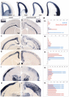
Fig. 1.
Fig. 1.
Cortical lamination abnormalities in Emx1/Ccm3 cKO neocortex. (A) Ccm3 mRNA is expressed in VZ/SVZ progenitors and newly postmitotic neurons migrating through the VZ/SVZ, but not in those migrating through the cortical plate at E13.5. (B) mRNA expression of Pax6 (radial glia), Tbr2 (SVZ progenitors) and Tbr1 (newborn neurons) in serial sections from the same embryo as in A. In situ hybridization as indicated. (C-L) In situ hybridization of brain sections of control (C,E,G,I,K) and Emx1/Ccm3 cKO (D,F,H,J,L) littermates. Emx1/Ccm3 cKO mutants lack clearly defined cortical layers: subplate neurons (Ctfg; C,D) are in the middle of the neocortex; L6 neurons (Tle4; E,F) are superficial to the subplate in a broad diffuse area; L5 neurons (Er81; G,H) are dispersed in upper cortex; L4 neurons (Rorb; I,J) are distributed in the upper half of the neocortical wall and L2-4 neurons (Cux2; K,L) in its entire depth. Control brains are Ccm3lox/lox (C and inset) or Emx1/Ccm3lox/+ (E,G,I,K). (C,D) P7; insets show high magnification of subplate neurons (P15); (E-L) P21. (M) The radial distribution of neurons expressing each marker is shown to the right of the respective in situ hybridization panels. Bars with broken lines indicate that all neurons in the bin are labeled and could not be counted. In Emx1/Ccm3 cKO neocortex, early-born neurons are shifted towards more superficial positions (bins 1-5), whereas late-born neurons are distributed across the neocortical wall (bins 1-10); many remain in deep positions (bins 6-10). Scale bars: 0.5 mm.
Fig. 2.
Fig. 2.
Migration defects of upper and deep layer cortical neurons in Ccm3 cKO mutants. (A-L) In situ hybridization of brain sections of control (A-C,G-I) and Ccm3 cKO mutant littermates [(D,J) hGfap/Ccm3 cKO; (E,K) Emx1/Ccm3 cKO; (F,L) NEX/Ccm3 cKO] with markers of deep [Er81 (A-F)] and upper [Cux2 (G-L)] layers at ages indicated. (A-F) L5 neurons are displaced superficially in Emx1/Ccm3 cKO (E), but at deep positions in hGfap/Ccm3 cKO (D) and NEX/Ccm3 cKO (F) mutants, comparable with controls (A-C). (G-L) Subsets of L2-4 neurons reside in ectopic deep positions in hGfap/Ccm3 cKO (J) but in upper cortex in NEX/Ccm3 cKO (L) mutants, comparable with controls (G-I). In Emx1/Ccm3 cKO, the majority of L2-4 neurons are in deep positions, indicating severely disrupted migration (K). Scale bar: 0.5 mm.
Fig. 3.
Fig. 3.
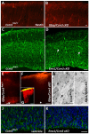
Radial glia defects in Ccm3 cKO mutants. (A-D) Immunofluorescent staining of radial glia (nestin) reveals disorganized processes in Ccm3 cKO mutants (B,D) compared with controls (A,C). Radial glia processes contact the pial surface (A,C), but are irregular in hGfap/Ccm3 cKO (B, E16.5) and Emx1/Ccm3 cKO mutants (D, E14.5). Arrows in D indicate abnormally fasciculated axons. (E-G) DiI labeling of cortical radial glia. At E15.5 (E,F) DiI-labeled cell bodies can be visualized at the VZ in control (E, arrow) but not in hGfap/Ccm3 cKO (F) embryos (asterisk indicates the ventricular surface). At E13.5 (approximate onset of hGfap-Cre action) labeled radial glia fibers span the cortical wall (G). (H,I) Electron micrographs show radial glia fibers in upper cortical plate in Ccm3lox/+ (H, arrow), but not in Emx1/Ccm3 cKO littermates at E14.5 (I). (J,K) Immunofluorescent staining of radial glia (nestin) reveals normal arrangement at the ventricular surface of E14.5 Emx1/Ccm3 cKO (K) and control littermates (J). Scale bars: 20 μm in A-D; 50 μm in E-G; 10 μm in H,I; 20 μm in J,K.
Fig. 4.
Fig. 4.
Cell-autonomous function of CCM3 in neural progenitors. (A-C) In utero electroporation (IUE) in the neocortical wall of Ccm3lox/lox embryos (E14.5) with CAG-GFP (A) or CAG-Cre and CAG-GFP (B) plasmids, analyzed at E16.5. In the presence of Cre (B), migration of electroporated cells is delayed. (C) Quantification of radial distributions of GFP-expressing cells. In the presence of Cre, the distribution of labeled cells is shifted toward the VZ (bin 1 is at the ventricular surface and bin 10 at the MZ). Error bars represent s.e.m. Student’s t-test, *P<0.05. (D-J) IUE in the neocortical wall of Ccm3lox/lox embryos (E13.5) with CAG-GFP (D,G,H) or CAG-Cre and Cre-responsive GFP (Stop-GFP) (E,I,J) plasmids, analyzed at E16.5. Following recombination, only a few GFP-expressing cells (thus, Cre-expressing) are in the cortical plate (cp), indicating delayed migration (E,I), compared with cells expressing GFP only (D,G). Quantification of the radial distribution of GFP-expressing cells from the outer IZ (bin 1) to the MZ (bin 5) (F). Significantly more Cre-expressing cells remain deep; GFP-only-expressing cells are in superficial positions. Error bars represent s.e.m. Student’s t-test, *P<0.05. High magnification of the leading processes of cells that have migrated into the cortical plate; compared with GFP-only-expressing cells, a large proportion of which extend long processes toward the cortical surface (89.43±6.44% in upper cortical plate), the leading processes of Cre-expressing cells are shorter and irregular, with a significant fraction (36.33±8.7%) displaying very short processes (H,J). Insets in G,I are low magnification views of G,H and I,J. Scale bars: 0.05 mm in A,B,D,E; 20 μm in G-J; 0.2 mm in insets (G,I).
Fig. 5.
Fig. 5.
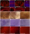
Abnormal cortical neuronal morphology in Ccm3 cKO mutants. (A-H) In utero electroporation (IUE) in the neocortical wall of Emx1/Ccm3 cKO and control littermates (E13.5) with the CAG-GFP plasmid, analyzed at E16.5 by fluorescence microscopy. (A,B) In control embryos (A), postmitotic neurons migrate into the cortical plate (cp) 3 days post-IUE; by contrast, many Emx1/Ccm3 cKO neurons remain in IZ (B). (C,D) In upper cortical plate, Emx1/Ccm3 cKO neurons are irregular and their leading processes do not reach the pial surface (D); control neurons are regularly arranged (C). (E,F) Migratory postmitotic neurons in upper IZ have transitioned into bipolar morphology (E; arrow indicates a leading process oriented towards the neocortical surface); however, Emx1/Ccm3 cKO neurons either remain multipolar or retain multiple short processes that are not oriented towards the pial surface [F; each of the four panels represents a different mutant neuron with distinct morphology; note multiple short processes (arrowheads)]. Neocortical surface is towards the top. (G,H) Low-magnification views of lateral neocortex showing axonal projections of electroporated neurons in control (G) and Emx1/Ccm3 cKO (H) littermates. Midline is towards the left. Axons extend towards the midline (G), but have irregular trajectories in the mutants (H). Insets are longer exposure views of axonal projections superimposed onto short exposure images of the electroporated hemisphere. (I-P) Morphology of primary cortical neurons. Immunofluorescent staining of neurons from Emx1/Ccm3 cKO (J,N) and hGfap/Ccm3 cKO (L,P) embryos, and control (I,K,M,O) littermates cultured for 3-5 days in vitro (DIV). (I,J) TUJ1 (I, control; J, Emx1/Ccm3Delta/lox); neurons isolated from E14.5 embryos at 5 DIV. (K,L) Falloidin staining of F-actin (K, control; L, hGfap/Ccm3 cKO); neurons isolated from E13.5 embryos at 5 DIV. (M,N) MAP2 (M: control; N: Emx1/Ccm3 cKO); neurons isolated from E14.5 embryos at 3 DIV. cKO neurons (N) develop multiple shorter dendrites when compared with controls (M). See also supplementary material Fig. S8G. (O,P) DCX (O, control; P, hGfap/Ccm3 cKO); neurons isolated from E15.5 embryos at 5 DIV. (Q-V) Golgi-Cox staining of adult Emx1/Ccm3lox/+ control (Q,S,T) and Emx1/Ccm3 cKO (R,U,V) brains. Dendritic trees are evident throughout the control neocortex, especially in upper layers (Q,S,T), but are underdeveloped in cKO (R,U,V). (Q,R) Symmetrical views of dorsomedial neocortex oriented towards the midline; (S,U) medial parietal cortex; (T,V) SSC. In all panels, the cortical surface is towards the top. Scale bars: 0.05 mm in A,B; 20 μm in C,D; 10 μm in E,F; 0.05 mm in G,H; 0.02 mm in I-P; 0.2 mm in Q,R; 0.05 mm in S-V.
Fig. 6.
Fig. 6.
Abnormal axonal trajectories in Ccm3 cKO mutants. (A-D) Immunofluorescent staining of axons (2H3) in control (A,B) and Emx1/Ccm3 cKO (C,D) or Emx1/Ccm3Delta/lox littermates (A-D: E14.5; E-H: E16.5). In dorsomedial mutant cortex, axonal bundles appear thinner (C) and aberrant axonal tracts are ectopically located (arrow in D) near the pia and abnormally fasciculated (D) compared with controls (B). Abnormalities in fasciculation and bifurcation of afferent and efferent axons (E-H). F,H are high-magnification views of E,G. (I-Q) The barrel field (I,L,O) is poorly formed in hGfap/Ccm3 cKO (J,M,P) and absent from Emx1/Ccm3 cKO (K,N,Q) mutants. Tangential sections through L4 of flattened P15 (I-K), P22 (L-N) or P8 (O-Q) cortices processed for cytochrome oxidase (CO) histochemistry (I-K), Nissl staining of postsynaptic L4 neurons (L-N) and immunofluorescent staining (vGlut2) of thalamocortical afferent terminals (O-Q). Scale bars: in A, 500 μm for A,C; in B, 20 μm for B,D; in E,F, 50 μm for E-H; in I, 200 μm for I-N; in O, 200 μm for O-Q.
Fig. 7.
Fig. 7.
RhoA activation in Ccm3 cKO mutant neocortex. (A) Western blot of neocortical protein extracts following a RhoA pull-down assay showing that activated Rho is elevated in hGfap/Ccm3 cKO neocortex at E15.5, compared with control samples. Total RhoA was used as loading control. Quantitative analysis indicates that activated RhoA is elevated 1.4-fold in hGfap/Ccm3lox/+ heterozygotes and 2.7-fold in hGfap/Ccm3 cKO mutants when compared with controls. Bars correspond to 25 kDa. (B) Histogram showing the quantification of the normalized signals obtained from A. Error bars represent s.e.m. (C) Western blot of neocortical protein extracts showing elevated ratio of inactive (Ser3 phosphorylated) cofilin to total cofilin in Emx1/Ccm3 cKO mutants. Quantitative analysis indicates that phospho-cofilin is increased 1.2-fold (E13.5; n=3) and 1.6-fold (P3; n=5) in Emx1/Ccm3 cKO mutants when compared with controls. Bars correspond to 25 kDa. (D) Histogram showing the quantification of the normalized signals obtained from (C). Error bars represent s.e.m. (E,F) Immunofluorescent staining of cortical neurons with phosphorylated cofilin shows more intense immunoreactivity in the neocortex of Emx1/Ccm3 cKO (D) when compared with control (C) (P3). Nuclei are counterstained with propidium iodide. Scale bar: 10 μm.
Fig. 8.
Fig. 8.
Summary of neuronal migration and positioning defects in Ccm3 cKO mutant neocortex. (A-F) Schematic drawings of the distribution of cortical pyramidal neurons in wild-type and Ccm3 cKO mice (P21). (A) Laminar positioning in wild-type mouse. (B) In hGfap/Ccm3 cKO mutants, cortical lamination is grossly normal. A subset of L2/3 neurons fails to migrate to the upper cortex. (C) In hGfap/Ccm3Delta/lox mutants, in which one Ccm3 allele is deleted in all cells and the other only in the radial glia lineage, most L5 neurons are displaced upwards near the pial surface; L4 neurons are distributed across the depth of the neocortex medially or displaced to the upper half laterally; upper layer neurons are dispersed across the neocortex. (D) In Emx1/Ccm3 cKO mutants, deep layer neurons are displaced upwards, whereas those of upper layers reside either across almost the entire depth of the neocortex (L4), or at its bottom two-thirds (L2/3); subplate neurons are confined to a diffuse median band. Most L2-4 neurons accumulate underneath L5/6. The relative positions of deep layer neurons are as in wild type, despite their ectopic location in upper cortex. (E) The Exm1/Ccm3Delta/lox mutants have a slightly more severe phenotype than the Emx1/Ccm3 cKO, reflected in a broad neuronal dispersion across all cortical layers. (F) Normal laminar positioning in NEX/Ccm3 cKO (and NEX/Ccm3Delta/lox, not shown) mutants. MZ, marginal zone; L, layer; SP, subplate; WM, white matter.

Similar articles

Cited by

References

    1. Alcamo E. A., Chirivella L., Dautzenberg M., Dobreva G., Fariñas I., Grosschedl R., McConnell S. K. (2008). Satb2 regulates callosal projection neuron identity in the developing cerebral cortex. Neuron 57, 364–377 - PubMed
    1. Angevine J. B., Jr, Sidman R. L. (1961). Autoradiographic study of cell migration during histogenesis of cerebral cortex in the mouse. Nature 192, 766–768 - PubMed
    1. Arlotta P., Molyneaux B. J., Chen J., Inoue J., Kominami R., Macklis J. D. (2005). Neuronal subtype-specific genes that control corticospinal motor neuron development in vivo. Neuron 45, 207–221 - PubMed
    1. Belvindrah R., Graus-Porta D., Goebbels S., Nave K. A., Müller U. (2007). Beta1 integrins in radial glia but not in migrating neurons are essential for the formation of cell layers in the cerebral cortex. J. Neurosci. 27, 13854–13865 - PMC - PubMed
    1. Bilgüvar K., Oztürk A. K., Louvi A., Kwan K. Y., Choi M., Tatli B., Yalnizoğlu D., Tüysüz B., Cağlayan A. O., Gökben S., et al. (2010). Whole-exome sequencing identifies recessive WDR62 mutations in severe brain malformations. Nature 467, 207–210 - PMC - PubMed

Publication types

MeSH terms

Substances

LinkOut - more resources