. 2014 Mar 3;9(3):e90858.
doi: 10.1371/journal.pone.0090858. eCollection 2014.

Effect of treadmill exercise timing on repair of full-thickness defects of articular cartilage by bone-derived mesenchymal stem cells: an experimental investigation in rats

Affiliations

Effect of treadmill exercise timing on repair of full-thickness defects of articular cartilage by bone-derived mesenchymal stem cells: an experimental investigation in rats

Jin-qi Song et al. PLoS One. .

Abstract

Objective: Current medical practice for the treatment of articular cartilage lesions remains a clinical challenge due to the limited self-repair ability of articular cartilage. Both experimental and clinical researches show that moderate exercise can improve articular cartilage repair process. However, optimal timing of moderate exercise is unclear. We aimed to evaluate the effect of timing of moderate treadmill exercise on repair of full-thickness defects of articular cartilage.

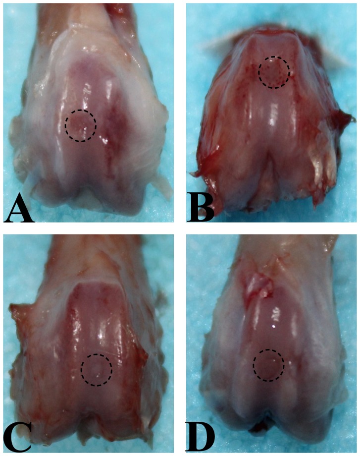
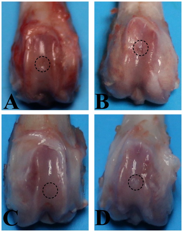
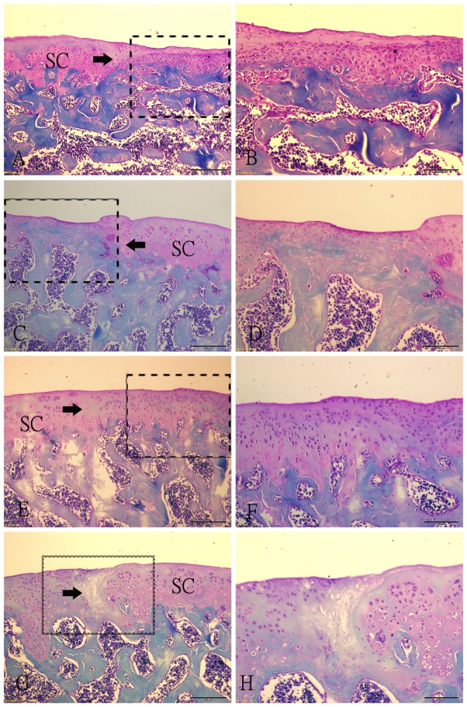
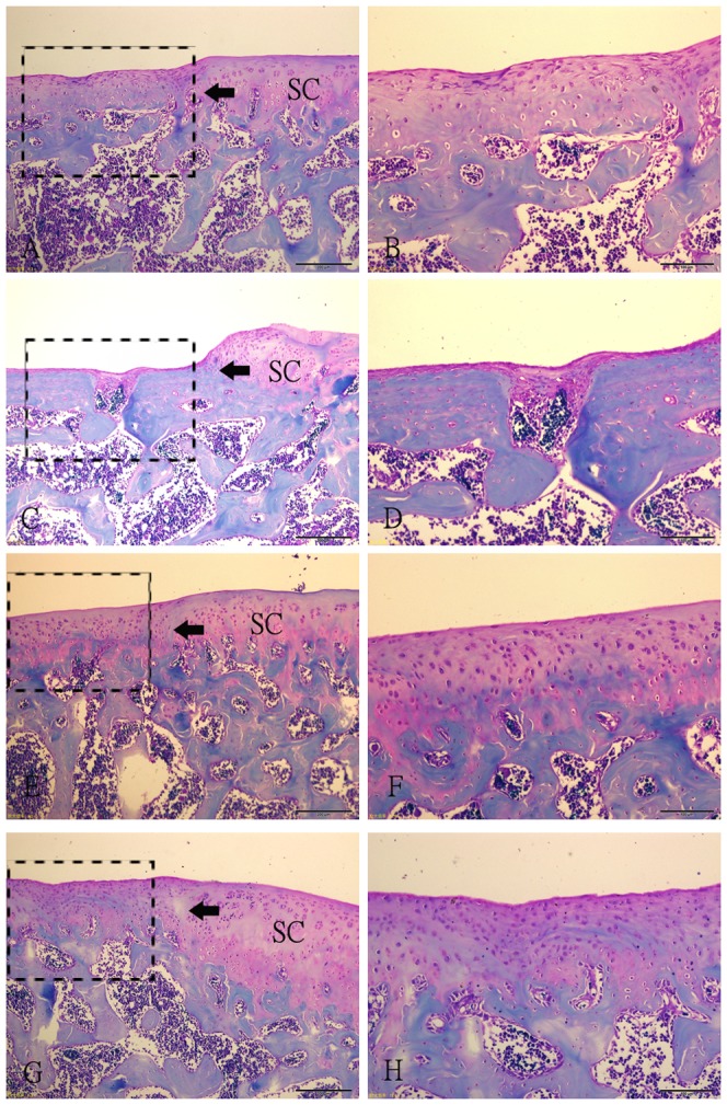
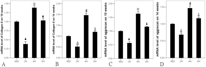
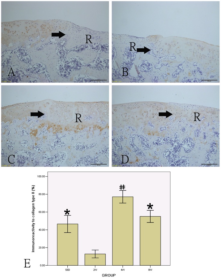
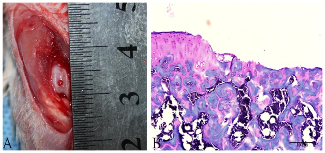
Design: Full-thickness cartilage defects were drilled in the patellar groove of bilateral femoral condyles in a total of 40 male SD rats before they were randomly assigned into four even groups. In sedentary control (SED) group, no exercise was given; in 2-week (2W), 4-week (4W) and 8-week groups, moderate treadmill exercise was initiated respectively two, four and eight weeks after operation. Half of the animals were sacrificed at week 10 after operation and half at week 14 after operation. Femoral condyles were harvested for gross observation and histochemical measurement by O'Driscoll scoring system. Collagen type II was detected by immunohistochemistry and mRNA expressions of aggrecan and collagen type II cartilage by RT-PCR.

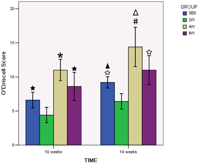
Results: Both 10 and 14 weeks post-operation, the best results were observed in 4W group and the worst results appeared in 2W group. The histochemistry scores and the expressions of collagen type II and aggrecan were significantly higher in 4W group than that in other three groups (P<0.05).

Conclusions: Moderate exercise at a selected timing (approximately 4 weeks) after injury can significantly promote the healing of cartilage defects but may hamper the repair process if performed too early while delayed intervention by moderate exercise may reduce its benefits in repair of the defects.


Conflict of interest statement

Competing Interests: The authors have declared that no competing interests exist.

Figures

Figure 1
Figure 1. Macroscopic appearance of an operated knee at explantation after 10 weeks of follow-up.
A. SED group. Defect is partially filled with repair tissue. B. 2W group. The margin of the normal cartilage around defect was eroded. C. 4W group. Defect is completely filled with white cartilage-like repair tissue to the level of surrounding uninjured cartilage, and the junction area is obvious. D. 8W group. Defect is filled with a white cartilage-like tissue with an irregular surface, and there is transparent line in the boundary area between the repair tissue and the residual cartilage.
Figure 2
Figure 2. Macroscopic appearance of an operated knee at explantation after 14 weeks of follow-up.
A. SED group. Defect is partially filled with repair tissue. B. 2W group. The margin of the normal cartilage around defect was eroded. C. 4W group. Defect is covered by smooth, glistening white tissue that was almost indistinguishable from the surrounding normal cartilage. D. 8W group. Defect is filled with a white cartilage-like tissue with an irregular surface, and the boundary of the defect was obvious.
Figure 3
Figure 3. Histological photographs of the representative repair tissues of defects in four groups at 10 weeks after operation with safranin-O staining.
(A–B) Fibrous tissue in SED group. The spindle-shaped fibroblasts embeded in thin fibrin-like tissue are present in the tissue. (C–D) Fibrous tissue in 2W group. The regenerative fibrous tissue is much thinner than that in SED group, and there are a little fibroblasts. The margin of the normal cartilage adjacent the defect is destroyed. (E–F) Repair tissue in 4W group. A large number of rounded cells embedded in the intensely Safranin-O stained extracellular matrix. The integration of the lesion to adjacent articular cartilage is good. (G–H) Regenerative tissue in 8W group. Some clusters of rounded cells resembling chondrocytes were recognized embedded in a fibrous and extracellular matrix. The bonding area is acellular. Fig. B, D, F, H (scale bar  =  200 µm) are higher magnifications of the junction of the repair tissue from Fig. A, C, E, G (scale bar  =  500 µm) respectively. SC: surrounding cartilage. →: defect margin.
Figure 4
Figure 4. O'Driscoll score in each group.
There was a significant increase in the 4W group compared to the to SED, 2W and 8W groups both in 10 weeks and 14 weeks after operation. * P<0.05 compared to SED, 2W and 8W group in 10 weeks after operation. #P<0.05 compared to SED, 2W and 8W group in 14 weeks after operation. ★ P<0.05 compared to 2W group in 10 weeks after operation. ☆ P<0.05 compared to 2W group in 14 weeks after operation. △P<0.05 compared to 4W group in 10 weeks after operation.▴P<0.05 compared to SED group in 10 weeks after operation.
Figure 5
Figure 5. Histological photographs of the representative repair tissues of defects in four groups at 14 weeks after operation with safranin-O staining.
(A–B) Fibrocartilaginous tissue in SED group. The extracellular matrix was poorly organized. (C–D) Fibrocartilaginous tissue in 2W group. The partly filled defect. The margin of the normal cartilage adjacent the defect is destroyed. (E–F) Repair tissue in 4W group. A large number of rounded cells embedded in the intensely Safranin-O stained extracellular matrix. The integration of the lesion to adjacent articular cartilage is good. (G–H) Regenerative tissue in 8W group. The surface of the repair tissue is irregular. The bonding area is acellular. Fig. B, D, F, H (scale bar  =  200 µm) are higher magnifications of the junction of the repair tissue and the residual cartilage from Fig. A, C, E, G (scale bar  =  500 µm) respectively. SC: surrounding cartilage. →: defect margin.
Figure 6
Figure 6. * P<0.05 compared to SED, 2W and 8W group in 10 weeks after operation.
#P<0.05 compared to SED, 2W and 8W group in 14 weeks after operation. ★ P<0.05 compared to SED group in 10 weeks after operation. Δ P<0.05 compared to SED group in 14 weeks after operation.
Figure 7
Figure 7. Immunohistochemical staining of collagen type II and the percentage of positive collagen type II in all groups (A: SED group; B: 2W group; C: 4W group; D: 8W group).
# P<0.05 compared to SED, 2W and 8W group; * P<0.05 compared to 2W group. (scale bar  =  100 µm). R: repair tissue. →: defect margin.
Figure 8
Figure 8. The full-thickness defects of patellofemoral articular surface.
A. Gross appearance of full-thickness articular surface defects. B. Safranin O-Fast Green staining of articular surface defects. (Original magnification × 200).

References

    1. Simon TM, Jackson DW (2006) Articular cartilage: injury pathways and treatment options. Sports Med Arthrosc 14(3): 146–154. - PubMed
    1. Widuchowski W, Lukasik P, Kwiatkowski G, Faltus R, Szyluk K, et al. (2008) Isolated full thickness chondral injuries. Prevalance and outcome of treatment. A retrospective study of 5233 knee arthroscopies. Acta Chir Orthop Traumatol Cech 75(5): 382–386. - PubMed
    1. Hurtig MB, Fretz PB, Doige CE, Schnurr DL (1988) Effects of lesion size and location on equine articular cartilage repair. Can J Vet Res 52(1): 137–146. - PMC - PubMed
    1. Kincaid SA, Van Sickle DC (1982) Effects of exercise on the histochemical changes of articular chondrocytes in adult dogs. Am J Vet Res 43(7): 1218–1226. - PubMed
    1. Zhuo Q, Yang W, Chen J, Wang Y (2012) Metabolic syndrome meets osteoarthritis. Nat Rev Rheumatol 8(12): 729–737. - PubMed

Publication types

LinkOut - more resources