. 2014 Mar 4;5(2):e00022-14.
doi: 10.1128/mBio.00022-14.

ZapE is a novel cell division protein interacting with FtsZ and modulating the Z-ring dynamics

ZapE is a novel cell division protein interacting with FtsZ and modulating the Z-ring dynamics

Benoit S Marteyn et al. mBio. .

Abstract

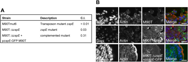
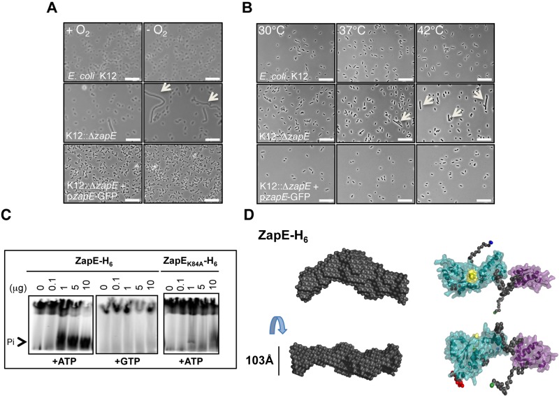
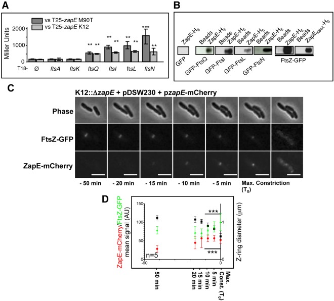
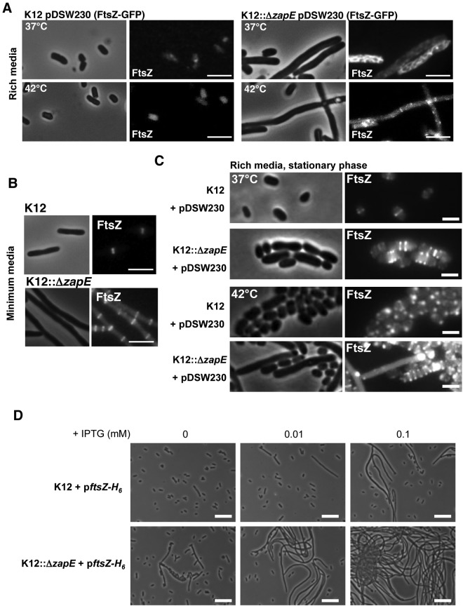
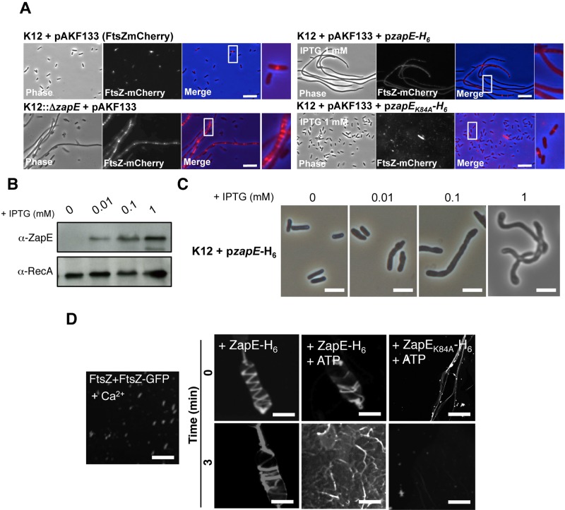
Bacterial cell division requires the formation of a mature divisome complex positioned at the midcell. The localization of the divisome complex is determined by the correct positioning, assembly, and constriction of the FtsZ ring (Z-ring). Z-ring constriction control remains poorly understood and (to some extent) controversial, probably due to the fact that this phenomenon is transient and controlled by numerous factors. Here, we characterize ZapE, a novel ATPase found in Gram-negative bacteria, which is required for growth under conditions of low oxygen, while loss of zapE results in temperature-dependent elongation of cell shape. We found that ZapE is recruited to the Z-ring during late stages of the cell division process and correlates with constriction of the Z-ring. Overexpression or inactivation of zapE leads to elongation of Escherichia coli and affects the dynamics of the Z-ring during division. In vitro, ZapE destabilizes FtsZ polymers in an ATP-dependent manner. IMPORTANCE Bacterial cell division has mainly been characterized in vitro. In this report, we could identify ZapE as a novel cell division protein which is not essential in vitro but is required during an infectious process. The bacterial cell division process relies on the assembly, positioning, and constriction of FtsZ ring (the so-called Z-ring). Among nonessential cell division proteins recently identified, ZapE is the first in which detection at the Z-ring correlates with its constriction. We demonstrate that ZapE abundance has to be tightly regulated to allow cell division to occur; absence or overexpression of ZapE leads to bacterial filamentation. As zapE is not essential, we speculate that additional Z-ring destabilizing proteins transiently recruited during late cell division process might be identified in the future.


Figures

FIG 1
FIG 1
ZapE is required during the Shigella infectious process in vivo. (A) Competitive index (C.I.) of a Shigella flexneri 5A zapE transposon mutant (M90Tmut6), a zapE mutant (M90T::ΔzapE), and a complemented strain (M90T::ΔzapE/pzapE-GFP M90T) in vivo. The C.I. assessed the ability of each mutant to colonize the rabbit ileal loop in comparison with the wild-type strain. A C.I. of 1 indicates no attenuation. Data represent averages of the results of three independent experiments. (B) Immunodetection of the M90T, M90T::ΔzapE, and M90T::ΔzapE/pzapE-GFP strains in the rabbit ileal loop model. DNA was stained with DAPI (blue) and actin with RRX-phalloidin (red). Shigella strains were labeled using α-LPS polyclonal antibody (pAb) (green). Image acquisition was performed using a confocal microscope. Right-hand panels show enlarged areas. Bars are 5 µm.
FIG 2
FIG 2
zapE inactivation leads to a stress-dependent (anaerobiosis, temperature) elongated phenotype of E. coli. zapE encodes an ATPase. (A) Anaerobiosis-dependent phenotype of E. coli wild-type, ΔzapE mutant, and complemented strains. Growth of K-12, K12::ΔzapE, and K12::ΔzapE/pzapE-GFP strains in minimum media in the presence (+O2) or absence (−O2) of oxygen at 37°C is shown. Scale bars are 10 µm. Arrows indicate elongated bacteria. (B) Temperature-dependent phenotype of K-12, K12::ΔzapE, and K12::ΔzapE/pzapE-GFP strains. Bacteria were grown in rich media at the indicated temperature until an OD600 = 0.5 was reached. Scale bars are 10 µm. Arrows indicate elongated bacteria. **, P < 0.01; *, P < 0.05. (C) ZapE or ZapEK84A ATPase activity assessed by silica layer chromatography. The reaction was performed in a Tris-HCl buffer (50 mM; pH 7.4) containing 10 mCi of radiolabeled ATPγ32 (or GTPγ32), 10 mM ATP, and 2.5 mM MgCl2 in the presence of various ZapE-H6 or ZapEK84A-H6 quantities, as indicated. (D) Most probable atom model of 15 DAMMIN reconstructions fitting the data at up to smax = 0.24 Å − 1 with an x = 1.683 and an NSD value of 0.7 ± 0.024, indicating the stability of the solution. The Walker A motif in the AAA+ ATPase is highlighted in yellow. N-terminal and C-terminal residues of ZapE are highlighted in green and blue.
FIG 3
FIG 3
ZapE is a cytoplasmic protein which interacts with FtsZ in vitro and in vivo. ZapE recruitment correlates with Z-ring constriction. (A) BACTH analysis was performed using the T25-zapE K-12 (E. coli K-12) or T25-zapE M90T (Shigella M90T) versus T18-plasmid constructs with indicated genes. Results are expressed in Miller units and were averaged from three independent experiments. Error bars show the standard deviations (SD). Comparing average activity to that of the T18 negative control, ** indicates P < 0.01 and *** indicates P < 0.001 (Student’s t test). (B) Pulldown assay with K-12 ZapE-H6 and GFP or GFP-tagged proteins in E. coli lysates. The interaction between E. coli FtsZ-GFP (pDSW230) and E. coli ZapE-H6 and ZapEK84A-H6, respectively, was analyzed using a His pulldown assay. (C) Expression and localization of FtsZ-GFP and ZapE-mCherry during a cell division. Time-lapse observation was performed on an LB-agar pad at 30°C, using a 200 M Axiovert epifluorescence microscope (Zeiss). Image acquisition was performed every 3 min (see also Movie S1 and Fig. S4C in the supplemental material for raw fluorescence quantification). This result is representative of five individual observations from three independent experiments. Bars are 2 µm. Max, maximum. (D) Relationship between, respectively, ZapE-mCherry and the FtsZ-GFP mean signal (AU) (K12::ΔzapE pzapE-mCherry) and the Z-ring diameter (pDSW230). Mean FtsZ-GFP and ZapE-mCherry fluorescent signals are represented in Fig. S4C in the supplemental material. n = 5 independent observations; error bars show the SD. *** indicates P < 0.001 (Student’s t test). Max. Const., maximum constriction.
FIG 4
FIG 4
Effect of ZapE inactivation on K-12 shape upon FtsZ-GFP and FtsZ expression modulation. (A) Localization of FtsZ-GFP (pDSW230) in K-12 and K12::ΔzapE strains grown in rich media (LB) at 37°C or 42°C in the absence of IPTG until an OD600 = 0.5 was reached. Bars are 5 µm. (B) Localization of FtsZ-GFP (pDSW230) in K-12 and K12::ΔzapE strains grown in minimum media (M9) at 37°C in the absence of IPTG until an OD600 = 0.5 was reached. Bars are 2 µm. (C) FtsZ-GFP (pDSW230) localization in K-12 and K12::ΔzapE strains during the stationary phase performed in LB rich media at 37°C or 42°C. These observations are representative of the results of at least three independent experiments. Bars are 1 µm. (D) Effect of FtsZ-H6 (WM971) overexpression on K-12 and K12::ΔzapE shape. Bacteria were grown in LB at 37°C in the presence of the indicated concentrations of IPTG until an OD600 = 0.5 was reached. These observations are representative of the results of three independent experiments. Bars are 10 µm.
FIG 5
FIG 5
ZapE level of expression perturbates Z-ring stability and bacterial shape. (A) Effect of zapE inactivation and ZapE or ZapEK84A overexpression on Z-ring stability (FtsZ-mCherry, pAKF133). Bacteria were grown in minimum media at 37°C in the presence of arabinose (0.01%) in addition to IPTG (1 mM) when indicated. Data are representative of the results of four independent observations (see also Fig. S5C in the supplemental material). (B) ZapE-H6 level of expression in strain K12-pzapE-H6 grown in LB at 37°C in the presence of the indicated concentrations of IPTG until an OD600 = 0.5 was reached. The level of expression of ZapE-H6 was assessed by Western blot analysis on bacterial whole extract using a polyclonal α-ZapE antibody. α-RecA was used as a control. (C) Phenotypic characterization of strain K12-pzapE-H6 grown under the conditions described for panel B. These observations are representative of the results of three independent experiments. Bars are 2 µm. (D) Polymerization of FtsZ (10 µM) with FtsZ-GFP (5 µM) in vitro was performed over 3 min, in the presence of 10 mM CaCl2, as described in references 16 and . ZapE-H6 or ZapEK84A-H6 (10 µM) was subsequently added to the reaction mixture (t = 0), with ATP added when indicated. Confocal imaging was performed at 0 and 3 min. Data are representative of the results of three independent experiments. Bars are 5 µm.

Similar articles

Cited by

References

    1. Bi EF, Lutkenhaus J. 1991. FtsZ ring structure associated with division in Escherichia coli. Nature 354:161–164. 10.1038/354161a0 - DOI - PubMed
    1. de Boer PA. 2010. Advances in understanding E. coli cell fission. Curr. Opin. Microbiol. 13:730–737. 10.1016/j.mib.2010.09.015 - DOI - PMC - PubMed
    1. Lutkenhaus J, Pichoff S, Du S. 2012. Bacterial cytokinesis: from Z ring to divisome. Cytoskeleton (Hoboken) 69:778–790. 10.1002/cm.21054 - DOI - PMC - PubMed
    1. Galli E, Gerdes K. 2010. Spatial resolution of two bacterial cell division proteins: ZapA recruits ZapB to the inner face of the Z-ring. Mol. Microbiol. 76:1514–1526. 10.1111/j.1365-2958.2010.07183.x - DOI - PubMed
    1. Hale CA, Shiomi D, Liu B, Bernhardt TG, Margolin W, Niki H, de Boer PA. 2011. Identification of Escherichia coli ZapC (YcbW) as a component of the division apparatus that binds and bundles FtsZ polymers. J. Bacteriol. 193:1393–1404. 10.1128/JB.01245-10 - DOI - PMC - PubMed

Publication types

MeSH terms